Fig. 5.
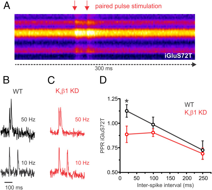
Paired-pulse measurements of glutamate release is impaired in Kvβ1 KD neurons. Optical measurements of glutamate release from individual presynaptic terminals expressing ultrafast iGluSnFR S72T during paired-pulse stimulation at various frequencies. (A) Kymograph of a representative recording across the axon of a hippocampal neuron recorded at 500 Hz and stimulated with a paired-pulse (50 Hz). Representative individual recordings of iGluSnFR S72T for a control (B) and Kvβ1 KD (C) neuron stimulated with 2APs at 50 Hz (Top) and 10 Hz (Bottom) normalized to the response of the first AP. (D) Average iGluSnFR paired-pulse ratio (normalized to glutamate reponse of first AP) from control and Kvβ1 KD neurons (control, n = 13 cells; Kvβ1 KD, n = 13 cells; *P < 0.05, Student’s t test; error bars represent SE). Note the selective impairment of release at 50 Hz compared to 10 and 4 Hz. Extracellular Ca2+ concentration is 2 mM in all experiments.

