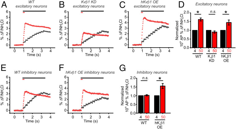
Fig. 6.
Synaptic facilitation is absent in Kvβ1 KD neurons. (A–C) Average traces of pHluorin measurements of exocytosis for WT (A), Kvβ1 KD (B), and human Kvβ1 overexpressed (C) excitatory neurons in responses to 10 APs delivered at 4 Hz (black) or 50 Hz (red) as measured with vG-pH. Bars at top of the graphs indicate the duration of each stimulation. (D) Normalization of average fusion induced by the 10th AP as a percentage of total vesicle fluorescences measured by application of NH4Cl. Neurons were stimulated with 10 AP at 4 Hz (black) or 50 Hz (red) (WT neurons, n = 16 cells; Kvβ1 KD neurons, n = 8 cells; hKvβ1 OE neurons, n = 6 cells; *P < 0.05, paired t test). (E and F) Average traces of pHluorin measurements of exocytosis for WT (E) and hKvβ1 overexpressed (F) inhibitory neurons in responses to 10 APs delivered at 4 Hz (black) or 50 Hz (red) as measured with vG-pH. Bars at top of the graphs indicate the duration of each stimulation. (G) Normalization of average fusion induced by the 10th AP as a percentage of total vesicle fluorescences measured by application of NH4Cl. Neurons were stimulated with 10 AP at 4 Hz (black) or 50 Hz (red) (WT neurons, n = 7 cells; hKvβ1 OE neurons, n = 5 cells; *P < 0.05, paired t test). Extracellular Ca2+ concentration is 2 mM in all experiments.

