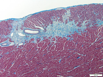
Fig. 2. Histopathologic image of the left ventricular wall in a North American river otter showing extensive areas of myofiber loss with replacement by fibrosis. Artery present displays segmental thickening of the tunica media by amorphous hyaline material or fibrosis. Masson’s trichome stain. Scale bar = 200 micrometers.

