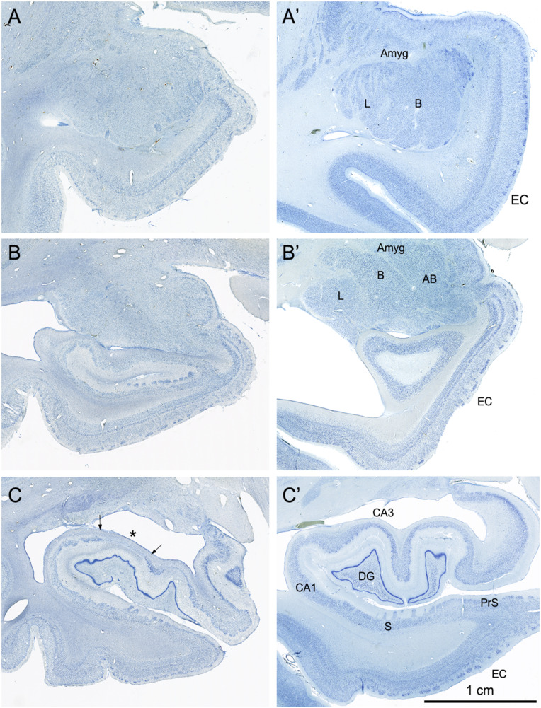
Fig. 4.
Coronal, Nissl-stained sections of the rostral, anterior, medial temporal lobe arranged from rostral (A) to caudal (C). All images are from the right hemisphere. (Left) Images from the brain of A.B. (Right) Images from the control comparison brain (IML-13) at approximately the same level. (A and A′) These sections illustrate a level through the rostral amygdala (Amyg) and demonstrate cell loss throughout. The entorhinal cortex (EC), located ventromedially, demonstrates patchy cell loss in A.B., particularly in layer III. (B and B′) This section is at a level through the caudal amygdala and at the rostral-most portion of the hippocampus. In the control section (B′), the lateral (L), basal (B), and accessory basal (AB) nuclei are easily differentiable. In A.B., there is generalized neuronal loss in the amygdala (B) which is replaced by a background of gliosis. The layers of the entorhinal cortex are apparent in A.B. but are overall less distinct due to patchy cell loss. (C and C’) This section is at the uncal level of the hippocampal formation and illustrates the rostral dentate gyrus (DG), hippocampus (CA3 and CA1), and components of the subicular complex (subiculum [S] and presubiculum [PrS]). Neurons of the polymorphic layer of the dentate gyrus are completely missing at this level as is the CA3 field of the hippocampus (asterisk between the two arrows in C). There is also neuronal loss in the deep portion of the CA1 pyramidal cell layer, which is better seen in Fig. 6).

