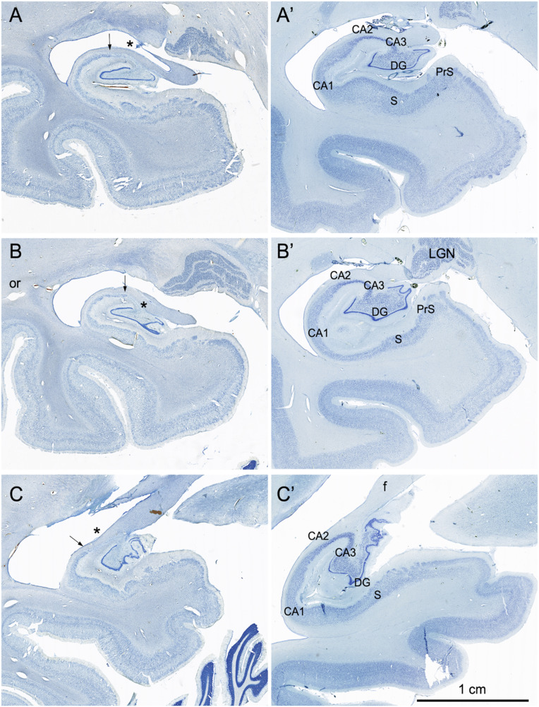
Fig. 5.
Coronal Nissl-stained sections of the medial temporal lobe of the right hemisphere arranged from rostral (A) to caudal (C). (Left) Images from the brain of A.B. (Right) Images from the control comparison brain (IML-13) at approximately the same level. The major loss of neurons in the sections from A.B. was in the CA3 region of the hippocampus (asterisk and up to arrow) and in the polymorphic layer of the dentate gyrus. (A and A′) Images of the hippocampal formation just caudal to the uncus. There are scattered cells at the arrow in a region containing the CA2 portion of the hippocampus. There is also cell loss in the deep portion of the pyramidal cell layer in the CA1 region of the hippocampus (better seen in Fig. 6). Some patchy cell loss is also observed in the subiculum (S). The presubiculum has a normal appearance. The parahippocampal region, like many other cortical areas, appears to be thinner than usual with patches of cell loss. (B and B′) Images of the hippocampal formation at the level of the caudal lateral geniculate nucleus (LGN). The pathology is similar to what was described in A. An expanded space in the region of the optic radiations (or) may correspond to the damage identified in an early CT scan of A.B. It may also be the cause of the patchy appearance of the cortex in V1 and adjacent visual areas (Fig. 9F) (C and C′) Images of the caudal pole of the hippocampal formation as the fimbria/fornix (f) ascends. The CA3 field of the hippocampus is completely devoid of pyramidal neurons (asterisk), and the polymorphic layer of the dentate gyrus is also totally depopulated of neurons.

