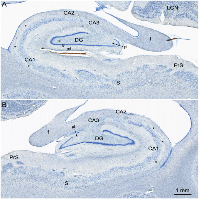
Fig. 6.
Higher magnification images of the right (A) and left (B) hippocampal formation in A.B. These sections are taken at a level through the rostral lateral geniculate nucleus (LGN). Here, it is clear that the granule cell layer (gl) of the dentate gyrus (DG) is irregular. Much of the polymorphic layer (pl) of the dentate gyrus is depopulated of neurons, but there are some scattered cells in the medial portion of the polymorphic layer (arrows). There appear to be remaining neurons in the CA2 field of the hippocampus (arrow with white arrowhead). It is also easy to appreciate that the deep portion of the CA1 pyramidal cell layer (asterisks) is devoid of neurons. The subiculum (S) and presubiculum (PrS) have an essentially normal appearance with occasional patches of cell loss. Additional abbreviation: ml, molecular layer of the dentate gyrus.

