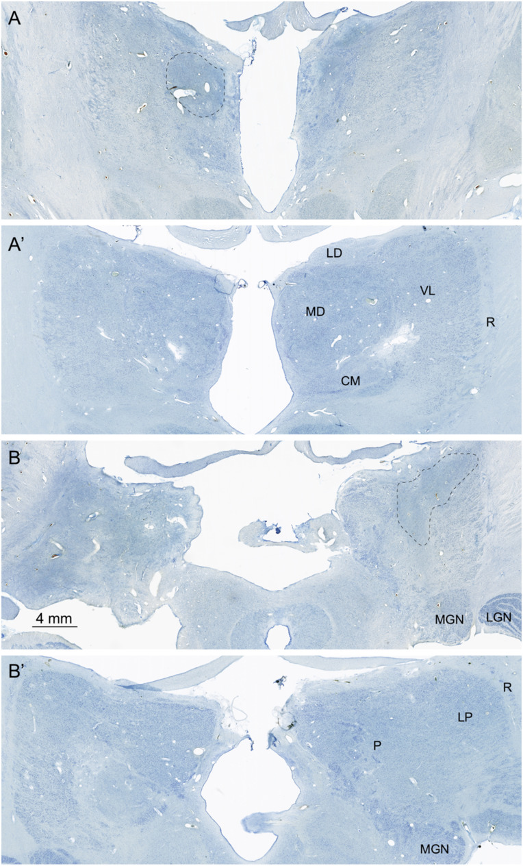
Fig. 7.
Nissl-stained images of the thalamus. (A and A′) This section is at a mid-rostrocaudal level with A showing A.B. and A′ showing a similar level from the control brain. There is substantial disruption of the structure of the thalamus in A.B. At this level, the mediodorsal (MD) nucleus is nearly entirely depopulated of neurons, and areas of gliosis (outlined region) are extensive. The ventral lateral (VL) nucleus located lateral to the mediodorsal nucleus is somewhat better preserved. The reticular nucleus (R) that surrounds the main portion of the thalamus cannot be distinguished in the section from A.B. (B and B′) These sections are taken through a caudal level of the thalamus that includes the pulvinar (P) and lateral posterior (LP) nuclei. The medial geniculate nucleus (MGN) can also be seen at this level. Virtually all of the pulvinar in A.B. is devoid of neurons, and there are large gliotic patches (outlined area). The medial (MGN) and lateral (LGN) geniculate nuclei of the thalamus are present in the section from A.B. and have a normal appearance. Additional abbreviations: CM, centre median nucleus of the thalamus; LD, lateral dorsal nucleus of the thalamus.

