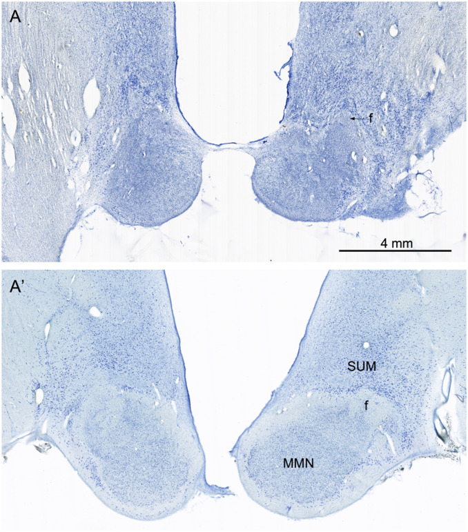
Fig. 8.
Nissl-stained sections from A.B. (A) and the control (A′) at a level through the main portion of the medial mammillary nucleus (MMN). In the control brain (A′), there are numerous neurons in the MMN. These are surrounded by a capsule of fibers that include a contribution from the incoming fornix (f) and collecting fibers that will contribute to the mammillothalamic tract. There are also larger and more darkly stained neurons located dorsal to the MMN that constitute the supramammillary region (SUM). In the section from A.B., the MMN is shrunken, and neurons are not detectable. The fibrous capsule is not obvious, and what appears to be the fornix is mainly gliotic. The neurons of the SUM are apparent but appear to be denser due to shrinkage of the lateral hypothalamic tissue. The arrow points to the shrunken fimbria in A.

