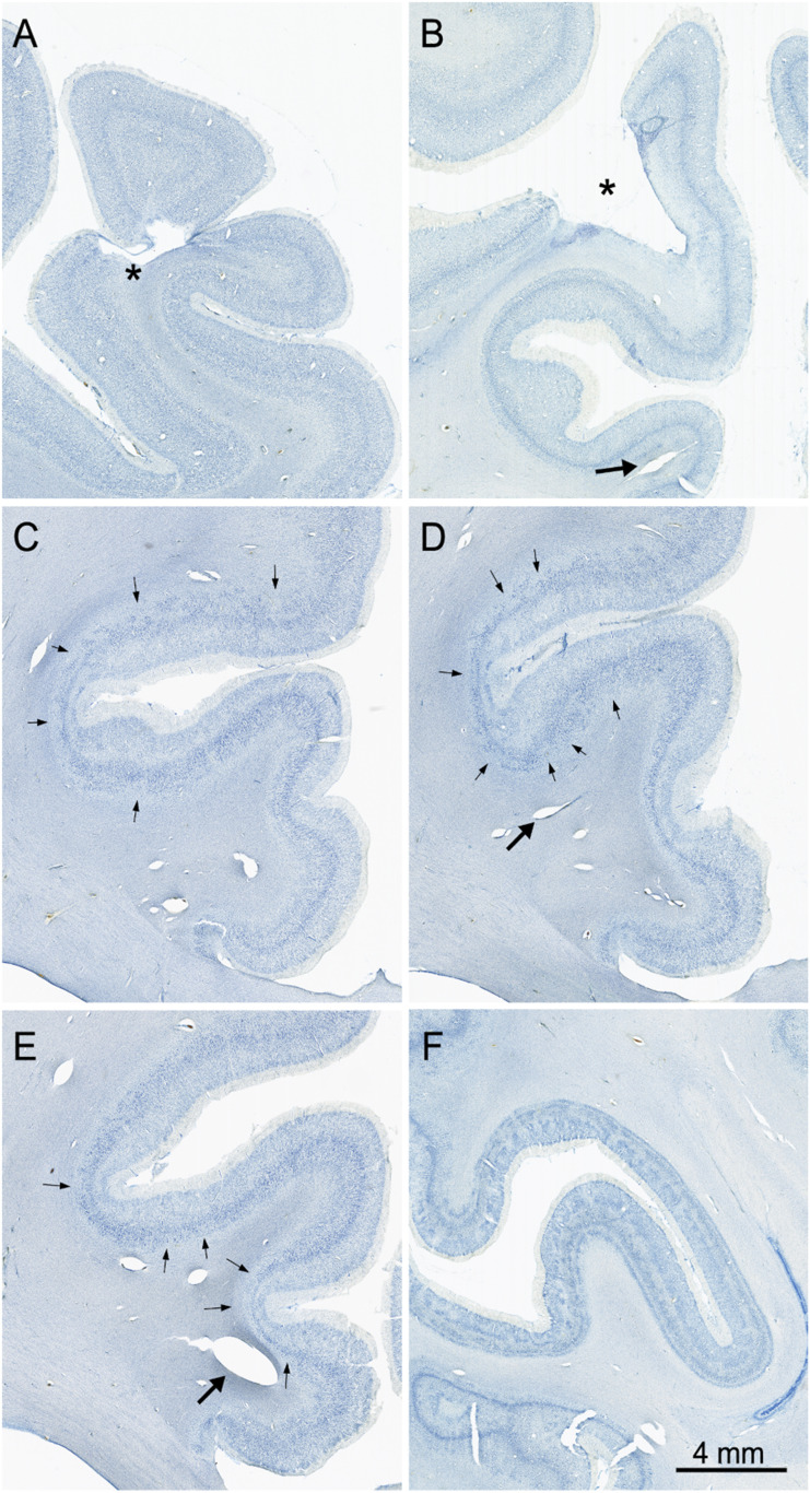
Fig. 9.
Images of Nissl-stained sections showing neuropathology in the cerebral cortex. (A) Infarct (*) in the left dorsomedial cortex. (B) Infarct (*) in the left posterior parietal cortex. (C–E) Sections arranged from rostral (C) through caudal (E) through the right cingulate cortex. Arrows point to patches of cell loss that occurred in all layers and was particularly consistent (and bilateral) in the cingulate cortex. Large arrows in panels B, C, and D point to expanded blood vessels in the white matter. (F) Radially oriented and laminar patches of cell loss in V1 cortex along both banks of the calcarine sulcus. The radial appearance of much of this pathology resembles ocular dominance columns. This may be due to damage to the optic radiations, which is observable in the right hemisphere (Fig. 5B).

