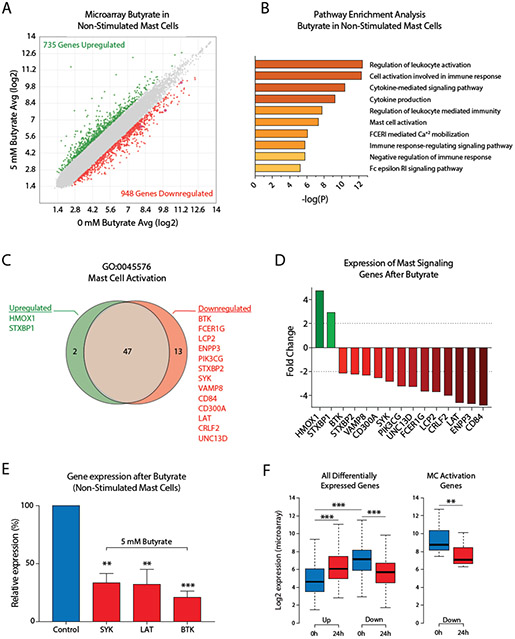
Figure 4. Butyrate induces human mast cell gene expression changes associated with cytokine signaling and mast cell activation.
A, Scatterplot comparison of gene expression levels measured by microarray analysis in unstimulated human mast cells either untreated or exposed to 5 mM butyrate for 24 h. Upregulated genes (>1.0 log2 fold change) are indicated in green, downregulated genes (<−1.0 log2 fold change) in red. B, Selected pathways that were strongly affected by butyrate treatment in unstimulated human mast cells. Y-axis denotes P-values on a −log10 scale. C, D, Butyrate-induced upregulation or downregulation of genes associated with the GO:0045576 – ‘Mast Cell Activation’ pathway represented as a Venn diagram (C) or bar graph (D) in unstimulated human mast cells treated with or without butyrate for 24 h. E, Butyrate-induced downregulation of BTK, SYK and LAT was validated using qPCR in human mast cells from 3 additional independent donors. F, Genes upregulated in response to 24h butyrate treatment (compared to vehicle treated cells, indicated as 0 h) showed overall low expression levels in vehicle treated mast cells, while downregulated genes showed significantly higher expression levels prior to treatment. Downregulated mast cell activation genes in particular are highly expressed before butyrate treatment (right boxplot). A-D, F, Results are from a single culture of human mast cells. E, Results are pooled from 3 independent experiments performed in human mast cells from 3 different donors. Data represent mean ± SEM, statistical significance was tested using a one-way ANOVA test: *P < 0.05; **P < 0.01; ***P < 0.001. GO = Gene ontology.

