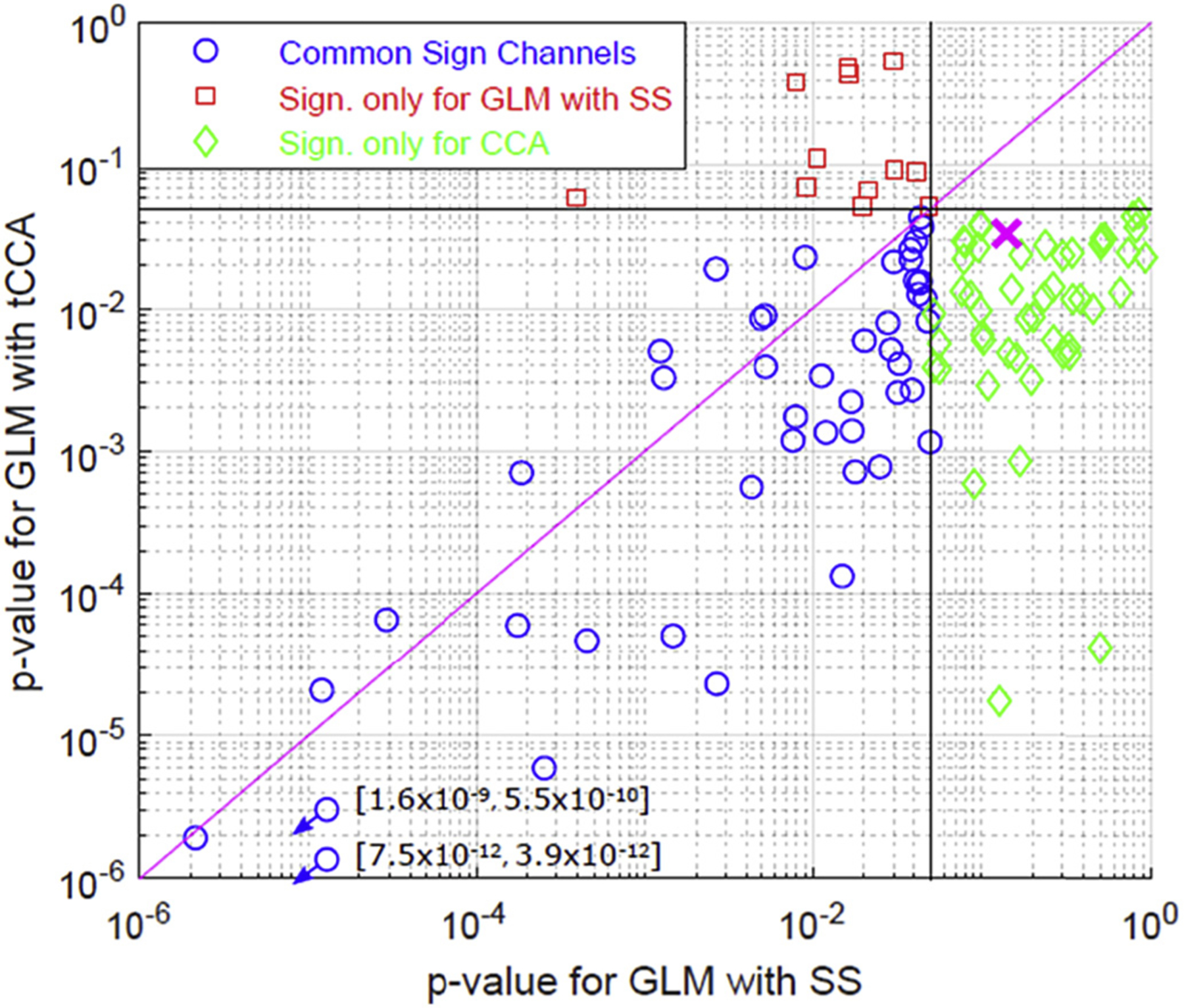
Fig. 11.

Comparison of active channels in visual stimulation data: Channels found significantly active only for GLM with tCCA (green) and only for GLM with SS (red). Blue circles show the channels that were found active with both methods. Magenta cross is the mean p-value for GLM with SS vs GLM with CCA across the combined set of channels that show significance using either of the methods.
