
Figure 4.
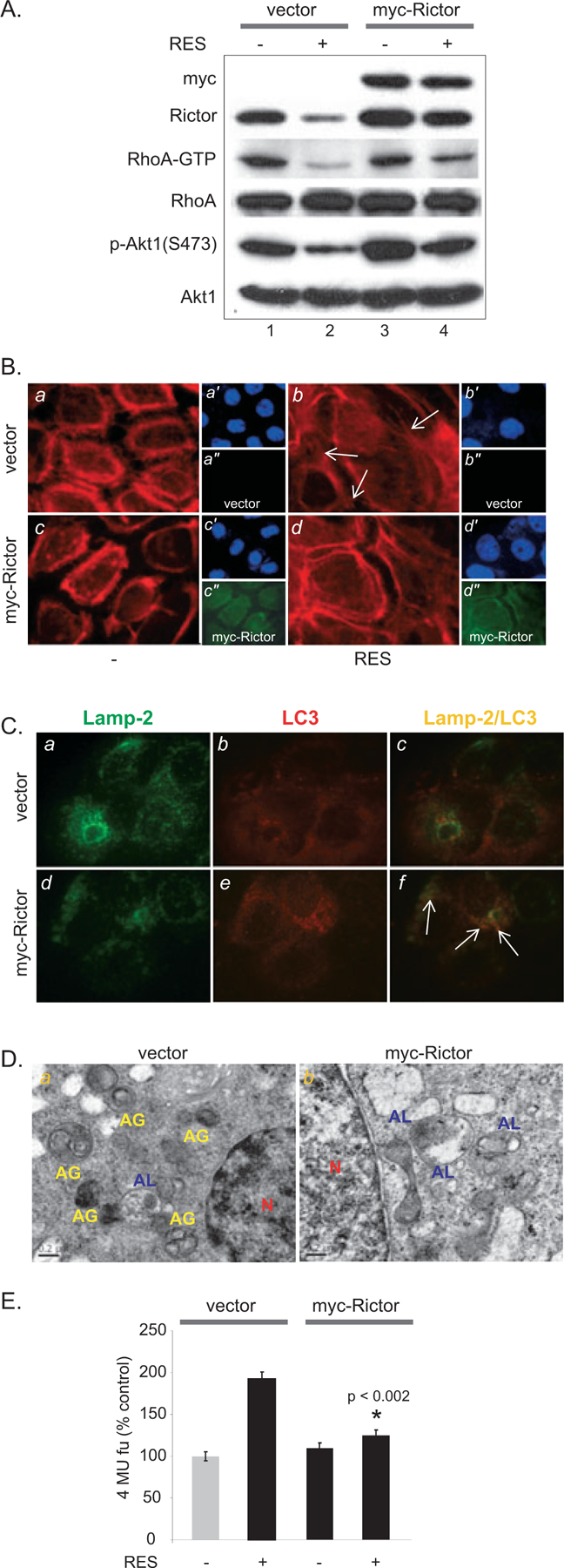
Exogenous Rictor expression restores RhoA-GTP activity and attenuates RES-induced SA-β-gal activity. (A) Exogenous Rictor restores RhoA-GTP activity. Cells were incubated with 50 μM RES for 48 h following viral transduction of myc-Rictor. (B) Actin remodeling is restored by exogenous Rictor expression in RES-treated A431 cells. F-actin was detected by rhodamine-phalloidin (red). Myc-Rictor expression is detected using anti-myc antibody (green, a″, b″, c″, d″). DAPI (blue, a′, b′, c′, d′). (C) Colocalization of Lamp-2 and LC3 is increased by exogenous Rictor expression. Green, Lamp-2; red, LC3, yellow, Lamp-2 ⁄ LC3. (D) EM images of A431 cells transduced with empty vector (a) or myc-Rictor (b) followed by RES treatment. AG, yellow, autophagosomes; AL, blue, autolysosomes; N, red, nucleus. (E) Rictor overexpression reduces RES-induced SA-β-gal activity.