. 2020 Nov 30;128(11):117009. doi: 10.1289/EHP7255

Figure 2.

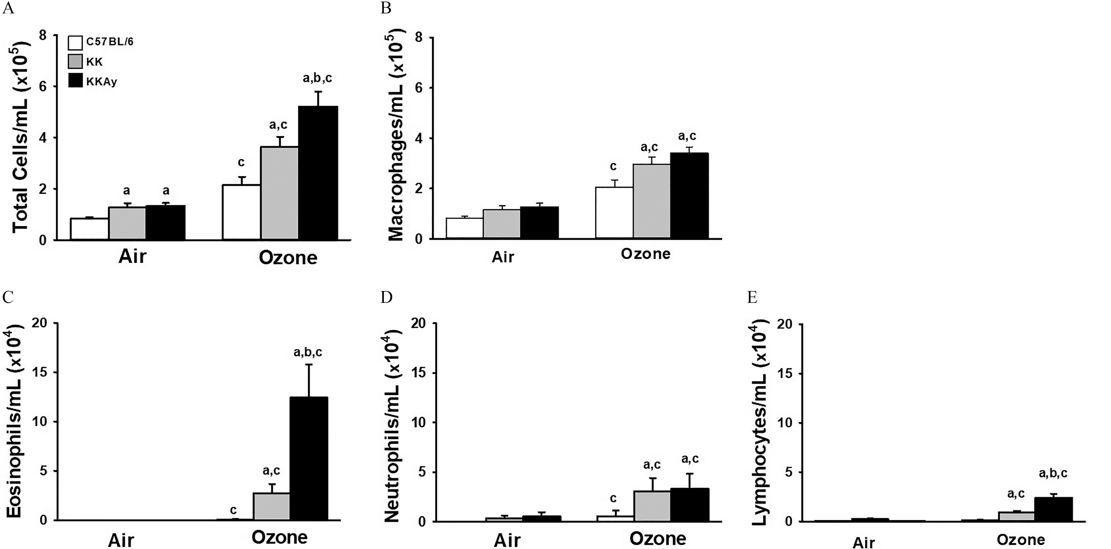
Figures 2A, 2B, 2C, 2D, and 2E are clustered bar graphs, plotting total cells per milliliter (times 10 begin superscript 5 end superscript), ranging from 0 to 8 in increments of 2, macrophages per milliliter (times 10 begin superscript 5 end superscript), ranging from 0 to 8 in increments of 2, eosinophils per milliliter (times 10 begin superscript 4 end superscript), ranging from 0 to 20 in increments of 5, neutrophils per milliliter (times 10 begin superscript 4 end superscript), ranging from 0 to 20 in increments of 5, and lymphocytes per milliliter (times 10 begin superscript 4 end superscript), ranging from 0 to 20 in increments of 5 (y-axis), respectively, across air and ozone (x-axis) for uppercase C 57 black 6, uppercase K K, and uppercase K K A y.

O3-induced bronchoalveolar lavage cellularity (BALF) cellularity in C57BL/6J, KK, and KKAy mice. Concentration in BALF of total cells (A), macrophages (B), eosinophils (C), neutrophils (D), and lymphocytes (E) were enumerated from cytospin preparations as described in “Methods.” Data are expressed as mean±SEM (n=78/group). (a) significantly different from similarly exposed C57BL/6 mice; (b) significantly different from similarly exposed KK mice; (c) significantly different from air-exposed mice of the same strain; p<0.05. Note: Data were analyzed using a completely randomized analysis of variance with factors of mouse strain and exposure, and comparisons of group means made with the Student–Newman–Keuls post hoc test. Summary data for panels A, B, C, D, and E can be found in Tables S11, S12, S13, S14, and S15, respectively.