. 2020 Nov 30;128(11):117009. doi: 10.1289/EHP7255

Figure 4.

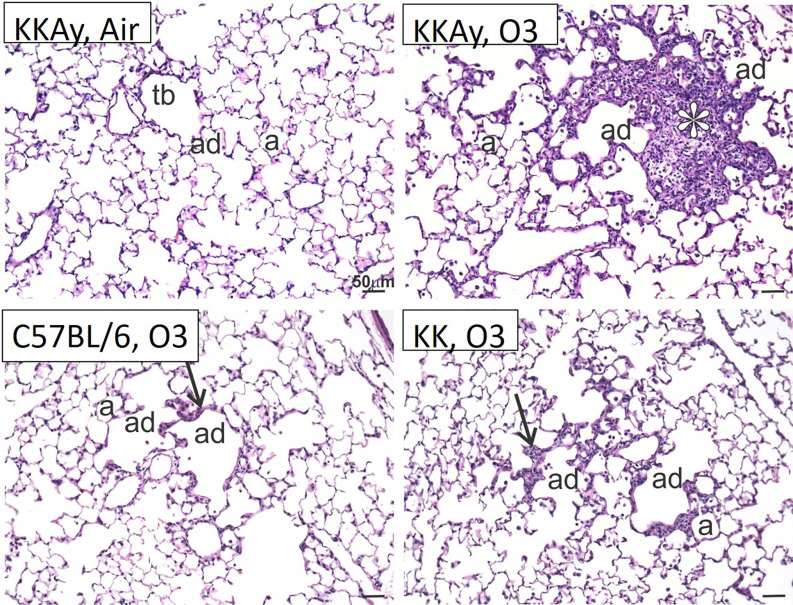
Figure 4 is a set of four light photomicrographs of centriacinar lesions, namely, K K A y after air, K K A y after ozone, uppercase C 57 black 6 after ozone, and K K after ozone. In the light photomicrographs, three tissues stained with hematoxylin and eosin, namely, terminal bronchiole, alveolar duct, and alveolus can be seen.

Histology of terminal airways in C57BL/6J, KK and KKAy mice. Light photomicrographs of centriacinar lesions (arrows, asterisk) in the lungs of KKAy, KK, and C57BL/6 mice exposed to O3. Tissues stained with hematoxylin and eosin. Note: Representative sections from the representative groups. a, alveolus; ad, alveolar duct; tb, terminal bronchiole.