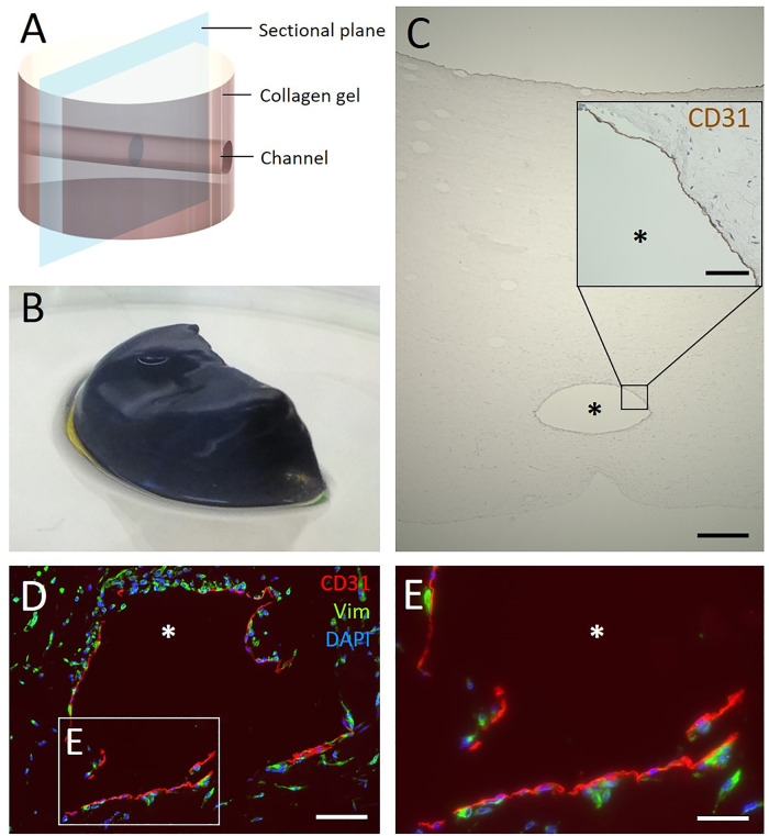
Fig 10. Analysis of the biofabricated hydrogel with channel.
(A) The schematic representation of the collagen hydrogel shows the position of the channels and the sectional plane for the following cross-sections. (B) Qualitative MTT-staining of half of a tissue construct. The blue color indicates viable cells within the hydrogel. (C) The central channel is visible in the HE-stained cross-section of the hydrogel after 14 days of culture in the bioreactor under dynamic flow conditions. The inset shows in detail the immunohistological staining of the channel. It reveals CD31-positive cells lining the channel lumen, indicating colonisation with endothelial cells. (D) Immunofluorescence staining of cross-sections of the channel visualizes the presence of endothelial cells and fibroblasts. Positive staining for CD31 (red) shows endothelial cells at the channel surface and positive staining for vimentin (green) shows fibroblasts within the hydrogel. Cell nuclei are labeled with DAPI (blue). (E) Magnification of D. The asterisk marks the channel lumen. Scale bars 500 μm (C), 100 μm (D) and 50 μm (inset in C, E).

