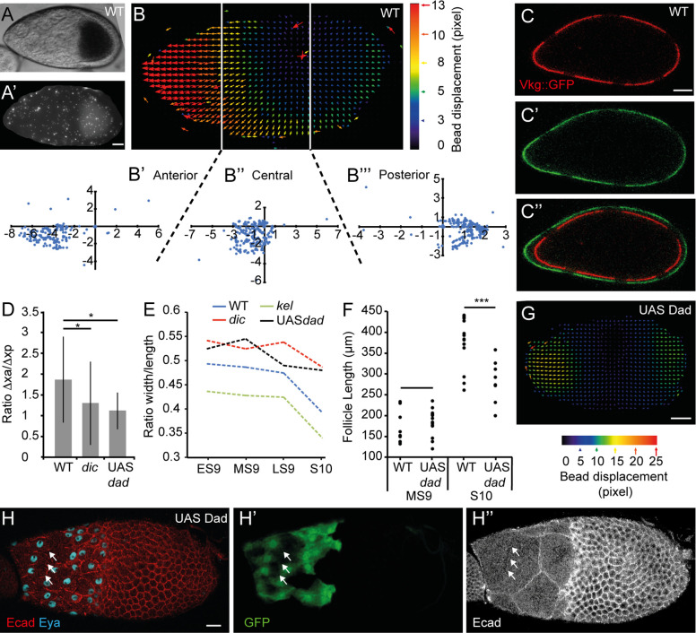
Fig 5. The follicles grow more anteriorly than posteriorly at S9.
(A) A WT MS9 follicle covered with fluorescent beads (A’). Beads outside the follicle were manually removed to simplify the analysis. (B) Particle image velocimetry representation from the beads positioned on the follicle shown in (A) and plots presenting the coordinates of the vectors for the anterior (B’), central (B”), and posterior (B”‘) areas. For each area, the initial (x, y) coordinates of the beads are (0,0). Each blue dot corresponds to the final x and y coordinates of a bead in μm. The vectors represent bead displacement (in pixel) and are color-coded as a function of their displacement. Duration of the experiment: 30 min. (C) WT MS9 follicle from a female carrying the vkg::GFP transgene to mark the BM (red in C, green in C’). Six bleached areas are visible at t = 0 (C) and at t = 80 min (C’). The two images have been overlaid by aligning the central bleached areas (C”) to show follicle growth during the interval. (D) Box and whisker plot of the ratio between the anterior (“xa”) and posterior (“xp”) growths of the BM in WT (n = 9), in dic follicles (n = 6), and in Dad-expressing follicles (n = 5). (E) Evolution of the ratio between the width and the length of the follicles from ES9 to S10 (n > 20 per stage). (F) Comparison of follicle length at MS9 and at S10B between WT and Dad-expressing follicles (n > 20 per stage). UAS-Dad is expressed under the tj-Gal4 driver. (G) Particle image velocimetry representation from the beads positioned on a Dad-expressing follicle. Duration of the experiment: 30 min. UAS-Dad is expressed under the tj-Gal4 driver. (H) MS9 Dad-expressing follicle. Only StC express Dad (GFP) (H’). The breakage of an NC membrane (arrows) are shown in H”. Scale bar: 20 μm. Data for graphs (D), (E), and (F) can be found in the S1 Data file. BM, basement membrane; dic, dicephalic; Ecad, Ecadherin; ES9, early S9; Eya, eye-absent expression; GFP, green fluorescent protein; kel, kelch; LS9, late S9; MS9, mid S9; S, stage; StC, stretched cell; WT, wild type.

