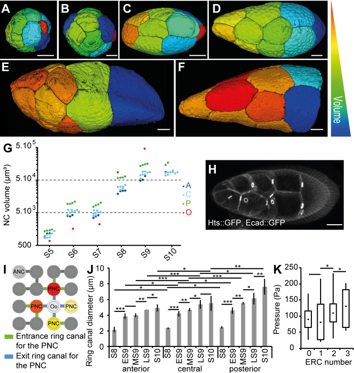
Fig 6. Intrinsic NC growth is important for the pressure gradient.
(A-F) The 3D reconstructions of germline cells in WT follicles at S5 (A), S6 (B), S7 (C), ES9 (D), LS9 (E), and S10 (F). Only NC, but not the Oo, are shown in (F). The arbitrary color scale indicates relative cell volumes for each follicle. (G) Individual volumes of anterior (“A,” dark blue), central (“C,” light blue), and posterior (“P,” green) NC and the Oo (red) from S5 to S10 WT follicles. For S10, the segmentation was incomplete. (H) An S9 follicle showing the RC (marked with Hts::GFP and visible as bright rings) between the NC. (I) Schematic representation of the 16 germline cells and their stereotypic connections through RC. The ANC is shown in light gray. The four PNCs (red, orange, and yellow) connect to the Oo (white) via the RC shown in blue. One PNC (light yellow) has no ERC, whereas the three others have one, two, or three ERCs (green). (J) Quantification of RC inner diameters from S8 to S10 as a function of their anterior (“A”), central (“C”), and posterior (“P”) localization (n = 5 per stage). (K) Box and whisker plot of inner pressure in the four PNCs as a function of the number of ERCs (n > 10 for NCs with zero, one, or two ERCs and n = 5 for NCs with three ERCs). Scale bar: 20 μm. Data for graphs (G), (J), and (K) can be found in the S1 Data file. ANC, anteriormost NC; Ecad, Ecadherin; ERC, entrance RC; ES9, early S9; GFP, green fluorescent protein; LS9, late S9; MS9, mid S9; NC, nurse cell; Oo, oocyte; PNC, posteriormost NC; RC, ring canal; S, stage.

