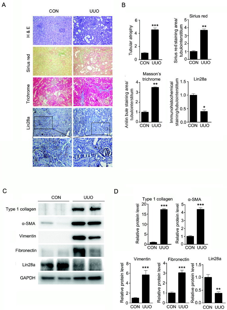
Fig. 1.
Effect of UUO-induced renal fibrosis on Lin28a expression and relative protein levels of type I collagen, α-SMA, vimentin, fibro-nectin and Lin28a in kidneys of mice with UUO. C57BL/6 mice were sacrificed 14 days after UUO. (A) Representative kidney tissue sections stained with hematoxylin and eosin (H&E), Sirius Red and Masson’s trichrome stain, and immunostained with antibody targeting Lin28a (Magnification, ×200). The number of atrophic tubules was determined by measuring the abnormal irregular and dilated tubular basement membranes in (H&E) stained kidney sections in five random fields under high-power magnification. Renal fibrosis area was assessed by Sirius Red and Masson’s trichrome staining. Lin28a expression in the tubulointerstitium of kidneys was measured by immunostaining. (B) Areas of positive staining with Sirius Red, Masson’s Trichrome and Lin28a in the UUO kidneys were quantitated by computer-based morphometric analysis and normalized to the control (=1) were expressed as the fold increase relative to the control in all bar graphs. Data are the mean ± SEM of five independent measurements (n = 5 in each group). *P < 0.05, **P < 0.01 and ***P < 0.001 compared with control mice. (C) Western blot analysis of protein levels of type I collagen, α-SMA, vimentin, fibronectin and Lin28a in the control and UUO kidneys. (D) Quantification of western blot analysis results expressed as the mean ± SEM of three independent measurements. GAPDH levels were analyzed as an internal control. **P < 0.01 and ***P < 0.001 compared with control mice.

