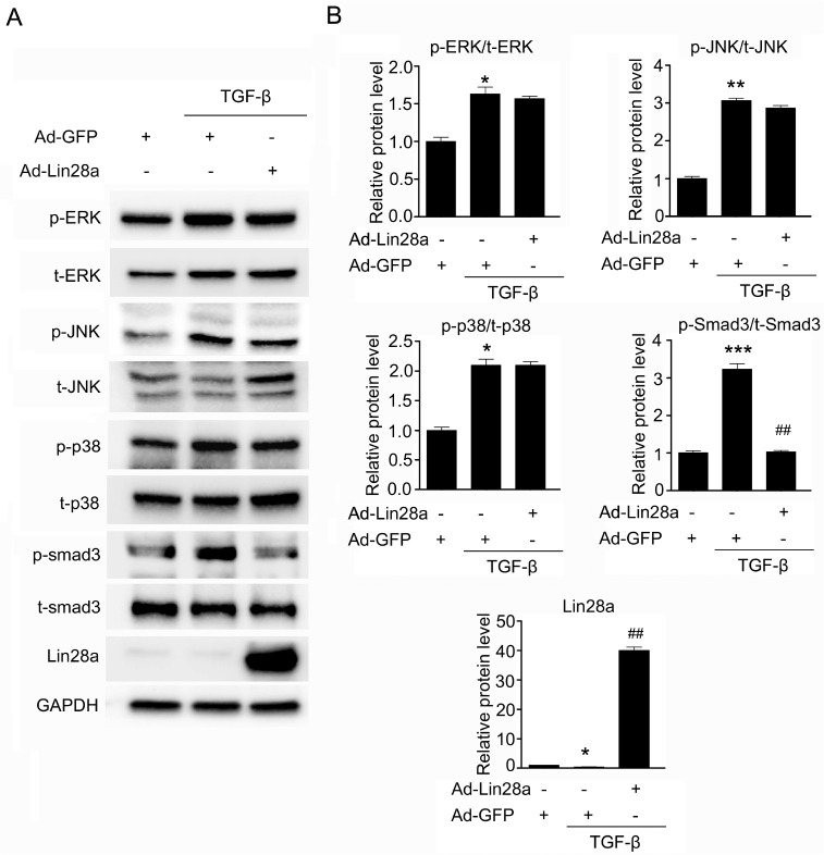
Fig. 4.
Effect of Lin28a on the TGF-β/SMAD3 signaling pathway. (A) Western blot analysis of the expression of p-ERK, p-JNK, p-p38 and p-smad3 in TGF-β-stimulated HK-2 cells. Cells were infected with 20 moi of Ad-Lin28a or Ad-GFP and then incubated with TGF-β (5 ng/ml) for 24 h. (B) Quantification of western blot analysis results expressed as the mean ± SEM of three independent measurements. GAPDH levels were analyzed as an internal control. *P < 0.05, **P < 0.01 and ***P < 0.001 compared with control, ##P < 0.001 compared with TGF-β alone.

