
FIGURE 1.
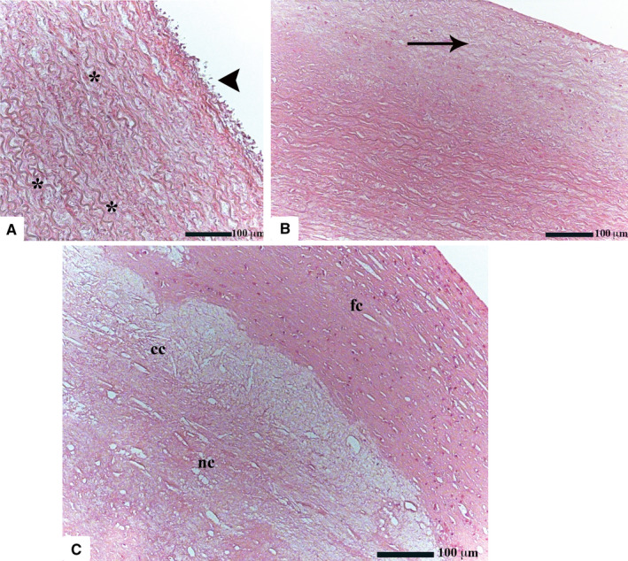
Histological features of healthy and atherosclerotic human aorta (H&E; original magnification ×50). Healthy ECs are very thin and flat (a). Their nuclei are frequently found to protrude into the lumen of the vessel (black arrowhead). In the tunica media, the characteristically wavy elastic fibers are entwined among collagenous fibers and SMCs. The aortic tissue adjacent to the lesion maintains a regular architecture showing only a mild intimal hyperplasia (black arrow) (b). Atherosclerotic plaques consist of a lipid‐rich necrotic core (nc) which is separated from the vascular lumen by a well‐developed fibrous cap (fc). Cholesterol crystals (cc), which appear as spindle‐shaped structures, can be also recognized (c)