
FIGURE 10.
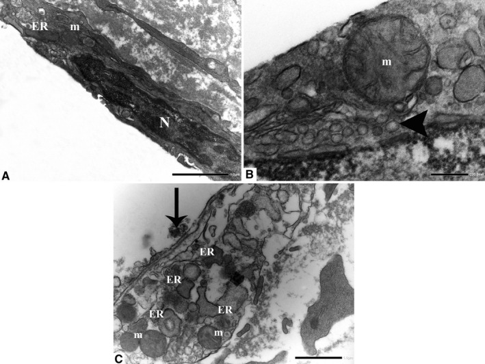
Endothelial cells with dilated ER membranes and altered mitochondria display cytoplasmic vacuolation (arrowhead). The nuclei frequently show areas of rarefaction and darker chromatin. An EC in an advanced stage of degeneration: Cell outlines are practically completely lost. Note the presence of dilated ER and altered mitochondria. ER: endoplasmic reticulum; m: mitochondria; and N: nucleus. (a, original magnification ×12000; b, original magnification ×40000; c, original magnification ×10000)