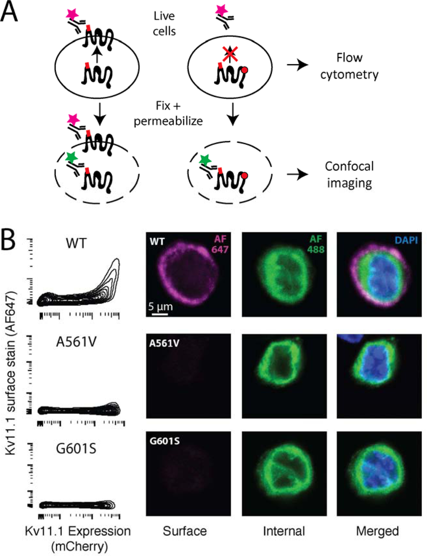
Figure 2: KV11.1 cell-surface trafficking assay.

A) Diagram of antibody labeling of surface or both surface and internal KV11.1. Live cells are stained with Alexa 647-labeled anti-HA antibody, which only labels surface KV11.1. Cells are assayed by flow cytometry. For confocal imaging, cells are fixed, permeabilized, and stained with an Alexa 488-labeled anti-HA antibody, which labels internal KV11.1. B) Flow cytometry assay of HEK293T cells expressing wildtype KV11.1, G601S (partial loss-of-function), or A561V (near total loss-of-function). The x-axis indicates mCherry level, which is a marker of protein expression. C) Confocal imaging of HEK293T cells expressing wildtype, G601S, or A561V KV11.1.
