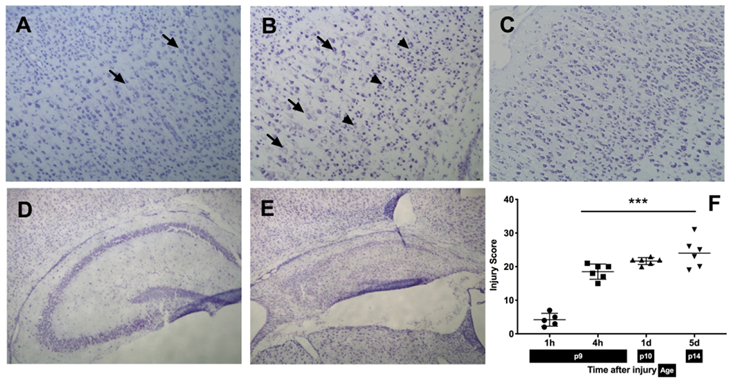
Figure 1:

A-E: Histopathological analysis of hypoxic-ischemic brain injury at different timepoints: cortex at 1 h (A) showing swelling of a few neuronal cells, 4 h (B) with increased area of swollen neurons (arrows), pyknotic cells (arrowheads) and 5 d (C) after the injury, showing the disruption in cortical structure and cell loss (10X magnification); hippocampus after HI at 4 h with no apparent injury (D) and 5 d (E) showing injured area with accumulation of shrunken, pyknotic cells and cystic infarction (4X magnification) F: Injury scores after HI at different timepoints: Injury extends from cortex at earlier timepoints to involvement of deep brain nuclei and adjacent cortex at the later timepoints.
