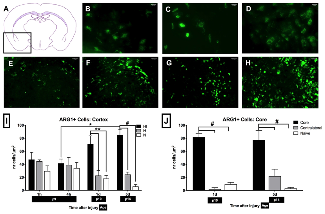
Figure 5: Changes in ARG-1+microglia after brain HI:

(A) Anatomical expression of ARG-1 in mice localizes to pyriform cortex, olfactory tubercle, pallidum, striatum, external capsule of corpus callosum, anterior commissure-temporal limb, amygdala, hypothalamus, caudoputamen. Change in ARG-1+ microglia morphology as a result of HI from bushy (B) into ameboid (C) and round phagocyting (D) phenotype. Changes in ARG-1+ microglia accumulation in striatum at different timepoints: N mice (E), and 4 h (F), 1 d (G) and 5 d (H) after injury. ARG-1+ microglia count increases in HI cortex (I) and HI core in striatum (J) at 1 d after injury and remains elevated for a prolonged time (n = 5-6 per HI and H group; n = 8 per N group, *p < 0.05, **p < 0.01, #p < 0.0001).
