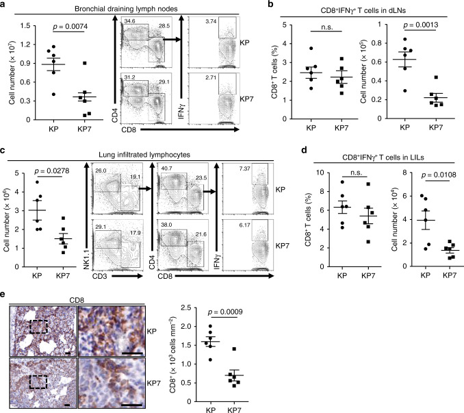
Fig. 4. CCL7 deficiency impairs CD8+ T cell expansion.
a Cell numbers and flow cytometry analysis of bronchial draining lymph nodes (dLNs) of KP (n = 6) and KP7 (n = 6) mice intranasally injected with Ad-Cre (1 × 10⁶) for 10 weeks. b Percentages and numbers of CD8+IFNγ+ T cells in bronchial dLNs from KP (n = 6) and KP7 (n = 6) mice treated as in a. c Lung infiltrated lymphocytes (LILs) of KP (n = 6) and KP7 (n = 6) mice injected with Ad-Cre (1 × 10⁶) for 10 weeks were isolated and calculated (left graph). LILs were stimulated with PMA and inomycin in the presence of Golgi-stop for 4 h followed by surface and intracellular staining with antibodies against NK1.1, CD3, CD4, CD8 and IFNγ and subject to flow cytometry analysis (right flow charts). d Percentages and numbers of CD8+IFNγ+ T cells in LILs from KP (n = 6) and KP7 (n = 6) mice treated as in a. e IHC staining (left images) and intensity or IOD analysis (right graph) of CD8 in tumor sections of KP (n = 6) and KP7 (n = 6) mice intranasally injected with Ad-Cre (1 × 10⁶) for 10 weeks. Two-tailed student’s t-test (a–e). n.s., not significant. Graphs show mean ± SEM (a–e). Scale bars, 50 μm. Data are representative of two independent experiments. Source data are provided as a source data file.

