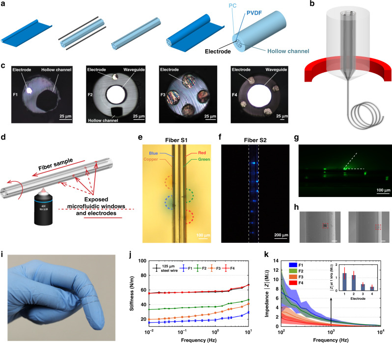
Fig. 1. Depth-dependent multifunctional fiber probe.
a A representative multifunctional fiber preform fabrication process (Fiber S1). b A schematic of the Fiber S1 thermal drawing process. c Cross-sectional images of fibers used in this study, electrode (BiSn). d A schematic of femtosecond laser micromachining process on Fiber S1. e–h Validation of the exposed microfluidic, optical excitation, and electrical windows: e four microfluidic windows were created on the four hollow channels of Fiber S1 and four different food colors were injected into the four channels respectively while the fiber was embedded in brain phantom; f an optical image showing the eight optical excitation sites fabricated on the eight waveguides of Fiber S2; g optical microscope image of the exposed Fiber S2 immersed in a drop of fluorescein excited by a 473 nm laser; h SEM images of the exposed microfluidic windows and electrodes (scale bar: 50 μm). i A photograph of functional fibers. j Bending stiffness measurements of Fiber F1–4. k Impedance measurements of the BiSn electrodes in Fiber F1–4. All error bars and shaded areas in the figure represent the standard deviation.

