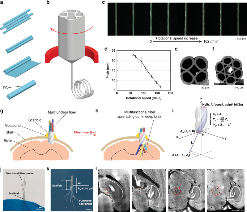
Fig. 2. Spatially expandable multifunctional fiber-based probes.
a Scaffolding fiber preform fabrication process. b Fiber drawing process with customized rotational feeding stage. c Side-view optical images of the scaffolding fibers drawn at different rotational speeds. d Plot showing the relatively linear relationship between the pitch and the rotational speed utilized in this study. e, f SEM images of the scaffolding fibers with five and seven hollow channels. g–i Schematics of the employment of the spatially expandable functional fiber probes: g scaffolding fiber is inserted into the brain and affixed by Metabond®; h functional fiber probes are further inserted into the brain tissue through the scaffolding fiber; i mathematical model of the locations of the inserted fiber probes. j Pre-validation of the expansion of the inserted functional fiber probes before implant surgeries. k Validation of the expandable fiber probes in the brain phantom. l Transverse DAPI-stained brain slices from the mouse with the spatially expandable fiber probes implanted for one week (n = 10). Representative images are shown taken from 10 animals. All error bars in the figure represent the standard deviation.

