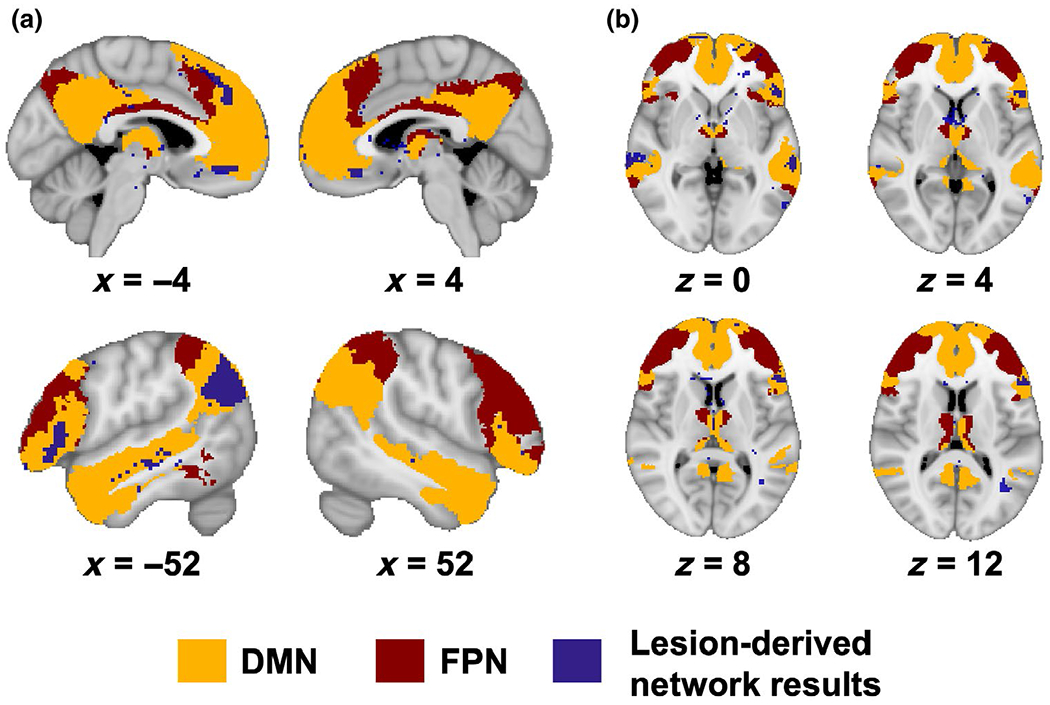
FIGURE 3.

Lesion network mapping results overlap with default mode and frontoparietal network masks. Thresholded voxel-based lesion network mapping results (p < 0.05, uncorrected; purple) are overlaid on masks of the default mode network (DMN) (orange) and frontoparietal network (FPN) (maroon) from a resting-state fMRI study of 1,000 participants (Yeo et al., 2011) and a study examining resting-state functional connectivity of thalamic nuclei major cortical networks (Hwang et al., 2017). (a) There is considerable overlap between the network results and regions in the DMN, and to a lesser extent with FPN regions as well as areas outside the DMN. (b) The network results intersect with thalamic nuclei functionally connected with the DMN and FPN. Results are displayed on the MNI-152 template
