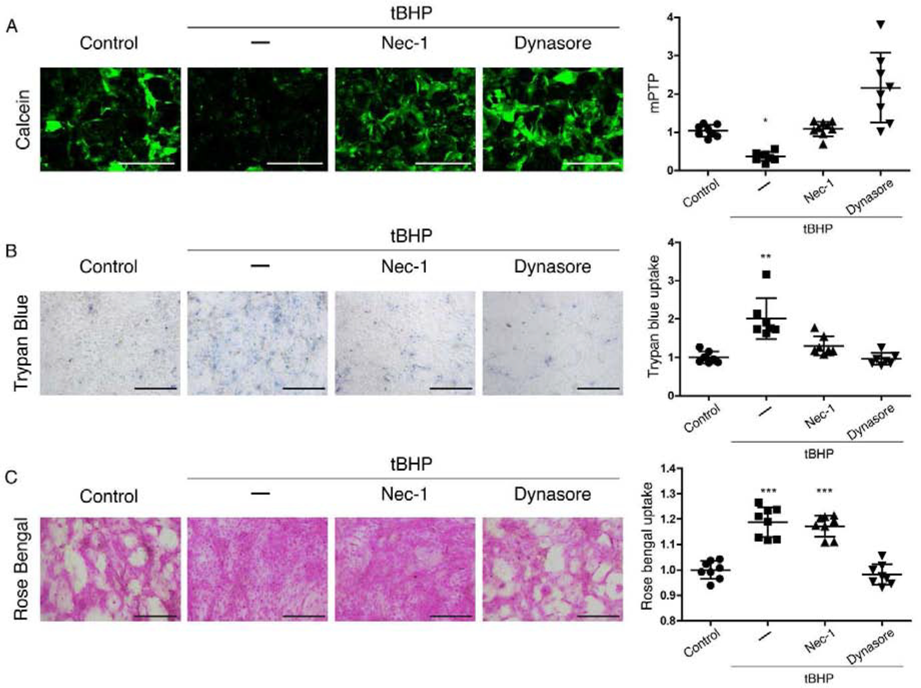
Figure 3. Dynasore and RIPK1 inhibitor necrostatin-1 protect mitochondria and the plasma membrane barrier in HCLE cells subjected to oxidative stress, but only dynasore protects the mucosal barrier.

Stratified cultures of HCLE cells with mucosal differentiation were exposed to tBHP (10 mM) for 2 hrs while being treated with either 300 μM necrostatin-1, 80 μM dynasore, or the same amount of DMSO vehicle (–). Unstressed cells kept in growth medium with the same DMSO concentration served as control.
(A) Representative images and quantification of mPTP opening, with the calcein-AM/CoCl2 assay (n = 3)
(B) Plasma membrane transcellular barrier integrity with trypan blue exclusion (n = 3)
(C) Mucosal transcellular barrier integrity with rose bengal exclusion (n = 3)
The data are presented as mean ± standard deviation. Significant differences were determined using the Kruskal-Wallis test with Dunn’s post-hoc test (A and B) or ANOVA with Bonferroni’s post-hoc test (C), after determining the homogeneity of the variances with the Bartlett’s test. * P<0.05; *** P<0.001. Scale bar: 1 mm (A); 200 μm (B, C).
