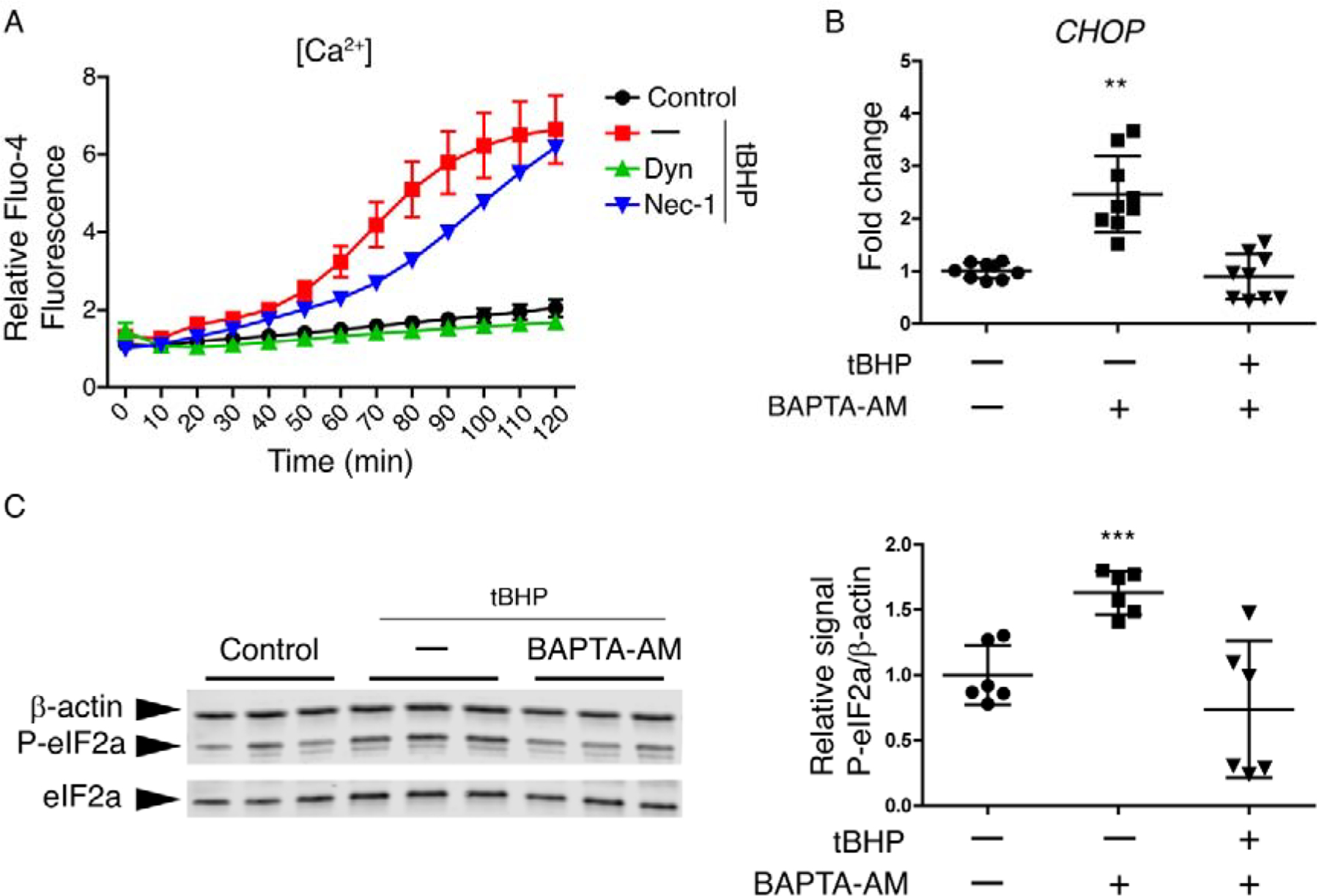
Figure 5. Dynasore inhibits the Ca2+ increase induced by oxidative stress, which is enough to reduce the activation of P-eIF2a/CHOP pathway.

(A) Monolayer cultures of HCLE cells were incubated with Fluo-4 Direct™ containing probenecid (2.5 mM). Then cells were exposed to tBHP (1 mM) for 2 hrs while being treated with either dynasore (40 μM), necrostatin-1 (300 μM), or DMSO vehicle (–). Unstressed cells kept in growth medium with the same DMSO concentration served as control. Changes in fluorescence over the times course were monitored under a fluorescence microscope. (n = 3)
(B and C) Stratified cultures of HCLE cells with mucosal differentiation were exposed to tBHP (10 mM) for 2 hrs after 1 h of preincubation with either BAPTA-AM (200 μM) or DMSO vehicle (–). Unstressed cells kept in growth medium with the same DMSO concentration served as control. At the end of the experiment, RNA or protein was isolated.
(B) Relative gene expression of CHOP was calculated with the 2−ΔΔCt method, using the levels of β-actin expression as housekeeping and the expression in control cells as the calibrator. n = 3.
(C) Representative images of western blot for P-eIF2α, eIF2α and β-actin and densitometry analysis of P-eIF2α normalized to β-actin levels as a loading control. n = 2.
The data are presented as mean ± standard deviation. Significant differences were determined using the Kruskal-Wallis test with Dunn’s post-hoc test. * P<0.05; ** P<0.01.
