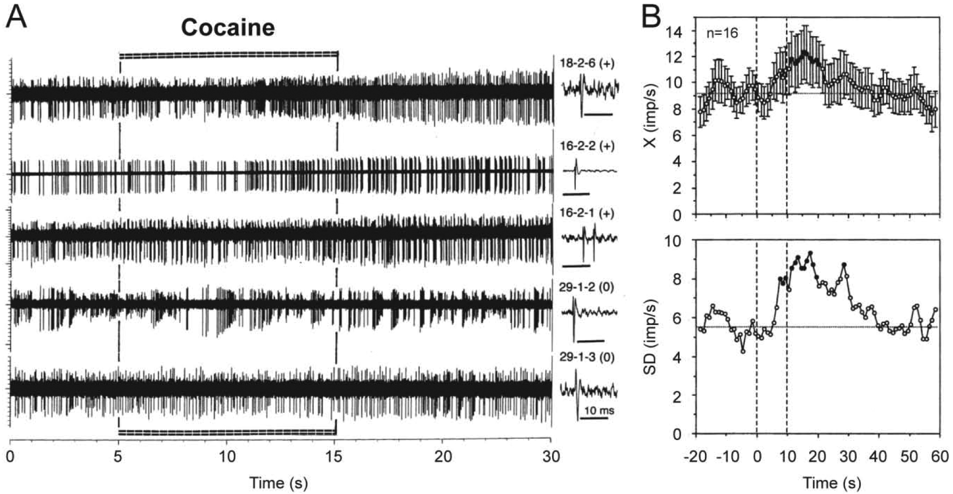
Figure 10. Changes in impulse activity of long-spike, presumed dopamine VTA neurons induced by iv cocaine at low reinforcing dose (0.25 mg/kg, 5–15 s) in freely moving rats.

A shows original records of neuronal responses for 20 s following cocaine injection and B shows mean (±SEM) changes in discharge rate (top; X, imp/s) and standard deviation of rate (bottom; SD, imp/s) for 60 s post-injection. Each original record was obtained from a different cell, whose numbers and single spikes are shown on the right panel of A. Note that three top traces show neuronal excitations (+) and the two lower traces show no changes in rate (0) but clear bursting. Mean data are shown for the entire group of tested cells, but 10 of 16 cells show excitations and 6 rats show no changes in rate of impulse activity. Filled symbols show values significantly different from baseline. Two vertical hatched lines on B at 0 and 10 s show the duration of cocaine injection. Original data were reported in Brown and Kiyatkin, 2008.
