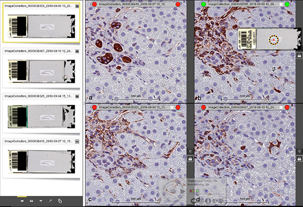
Figure 2.
Side-by-side (descending order) viewing of four different antibodies from smIHC using Leica SlidePath Digital Image Hub software. (a) K19, (b) vimentin. (c) αSMA and (d) S100A4. The four images can be synchronized in the same view pane by “locking” them together with the padlock icon. As a result, location and magnification changes made to one pane automatically apply to the other three panes. Scale bar = 100 μm.

