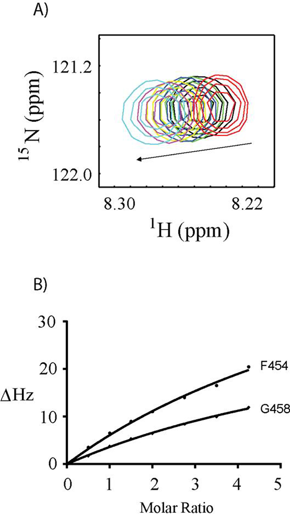
Figure 2. S100A10-AnxA2Nter titration to Ac-15N-F454G458G461-SW.
A: Chemical shift changes of F454 cross peak versus the titration of S100A10-AnxA2Nter fusion protein. The molar ratio between S100A10-AnxA2Nter fusion protein and Ac-15N-F454G458G461-SW was color coded as follows: Red 0:1; Black 1:1; Green 1.5:1; Blue 2:1; Yellow 2.75:1; Magenta 3.5:1 and Cyan 4.3:1. B: The curve fitting of chemical shift perturbation for residues F454 and G458 of Ac-15N-F454G458G461-SW titrated by S100A10-AnxA2Nter fusion protein. The CSP values in unit of HZ was calculated using the following equation. . The ΔωN and ΔωH are the nitrogen and proton chemical shift difference between free Ac-15N-F454G458G461-SW and that in the mixture with addition of S100A10-AnxA2Nter.

