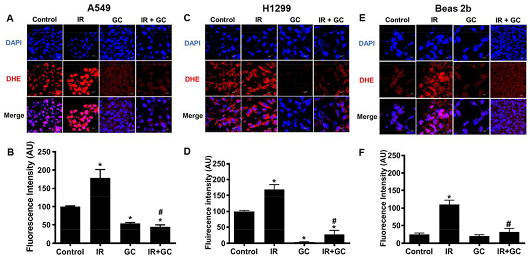
Fig. 6: Detection of superoxide with using the fluorescence probe DHE.

A549, H1299 and Beas 2b cells grown on a coverslip were either left untreated or treated with GC4419 (GC;10 μM) alone, IR (8Gy) alone or in combination, then maintained in a humidified atmosphere of 95% air: 5% CO2 at 37 0C. At 120 minutes post IR, the drug was washed out and attached cells were incubated for 30 minutes with DHE that reacts with superoxide giving rise to red fluorescence. Cells were imaged (A, C, E) by confocal microscopy. The quantitation of the images are shown in (B, D, F). GC4419 largely quenched the observed fluorescence confirming that it was superoxide-derived. The results of triplicate measurements are shown with mean ± SEM. * significant from control (p<0.001), # significant from IR (p< 0.001).
