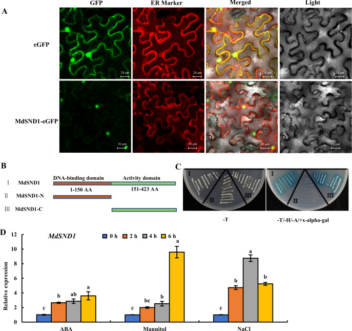
Fig. 2. Subcellular localization, transcriptional activation, and stress induction of MdSND1 in apple.
A Subcellular localization of pRI-MdSND1-eGFP in epidermal cells of tobacco leaves. B Amino acid sequence structure of full-length MdSND1, the MdSND1 N-terminal region and the MdSND1 C-terminal region. C Transcriptional activation assays of MdSND1. The GAL4 DNA-binding domain (GAL4DB) was fused to the MdSND1 N-terminal region, MdSND1 C-terminal region, and full-length MdSND1 sequence, after which the constructs were transferred into yeast. (−T/−H/−A+X-alpha-gal) means selective medium supplemented with 5-bromo-4-chloro-3-indolyl-alpha-D-galactoside but lacking Trp, His, and adenine, while (−T) indicates selective medium lacking Trp. D MdSND1 was induced by osmotic stress. The error bars indicate the standard deviations (SDs) of three biological replicates. The letters indicate the level of significance (P < 0.05, according to Duncan’s multiple range test)

