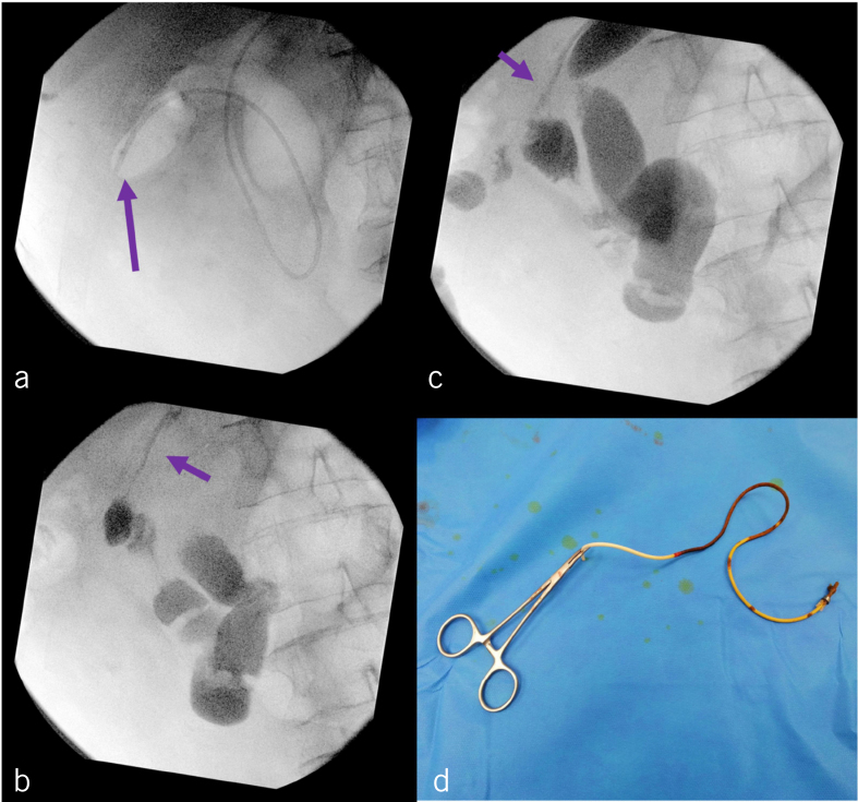
Figure 3.
(a) Intraoperative fluoroscopy showing the location of catheter in the small bowel (arrow). (b) Contrast placed through the catheter (arrow) to confirm the migrated catheter was in the small bowel rather than the hepatic artery. (c) Contrast placed through the fistula tract (arrow) to ensure no contract extravasation intraperitoneally after removal of the catheter. (d) The removed catheter in entirety.

