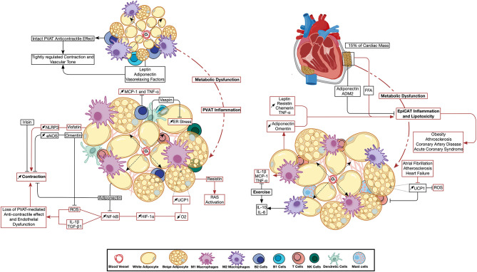
Figure 2.
Perivascular and epicardiac adipose tissue dysfunction: the emerging role of immune cell and adipokine profile dysregulation. Metabolic impairment triggers changes in PVAT (left) and EpiCAT (right) adipokine environment and immune cell activity. Inflammation has a direct effect on the neighboring vascular and cardiac tissue. Changes in UCP1 expression were reported to have opposite effects in either depot. Pathways active in basal PVAT and EpiCAT homeostasis are depicted in black arrows, while those activated during inflammation are shown in red. ADM2, Adrenomedullin-2; eNOS, Endothelial Nitric Oxide Synthase; EpiCAT, Epicardial Adipose Tissue; ER Stress, Endoplasmic Reticulum Stress; HIF-1α, Hypoxia-induced Factor 1 Alpha; IL, Interleukin; MCP-1, Monocyte Chemoattractant Protein 1; NF-κB, Nuclear Factor Kapp-light-chain-enhancer of Activated B cells; NLRP3, NLR Family Pyrin Domain Containing 3; O2, Oxygen; PVAT, Perivascular Adipose Tissue; RAS, Renin Angiotensin System; ROS, Reactive Oxygen Species; TGF-β, Transforming Growth Factor Beta; TNFα, Tumor Necrosis Factor Alpha; UCP1, Uncoupling Protein 1.

