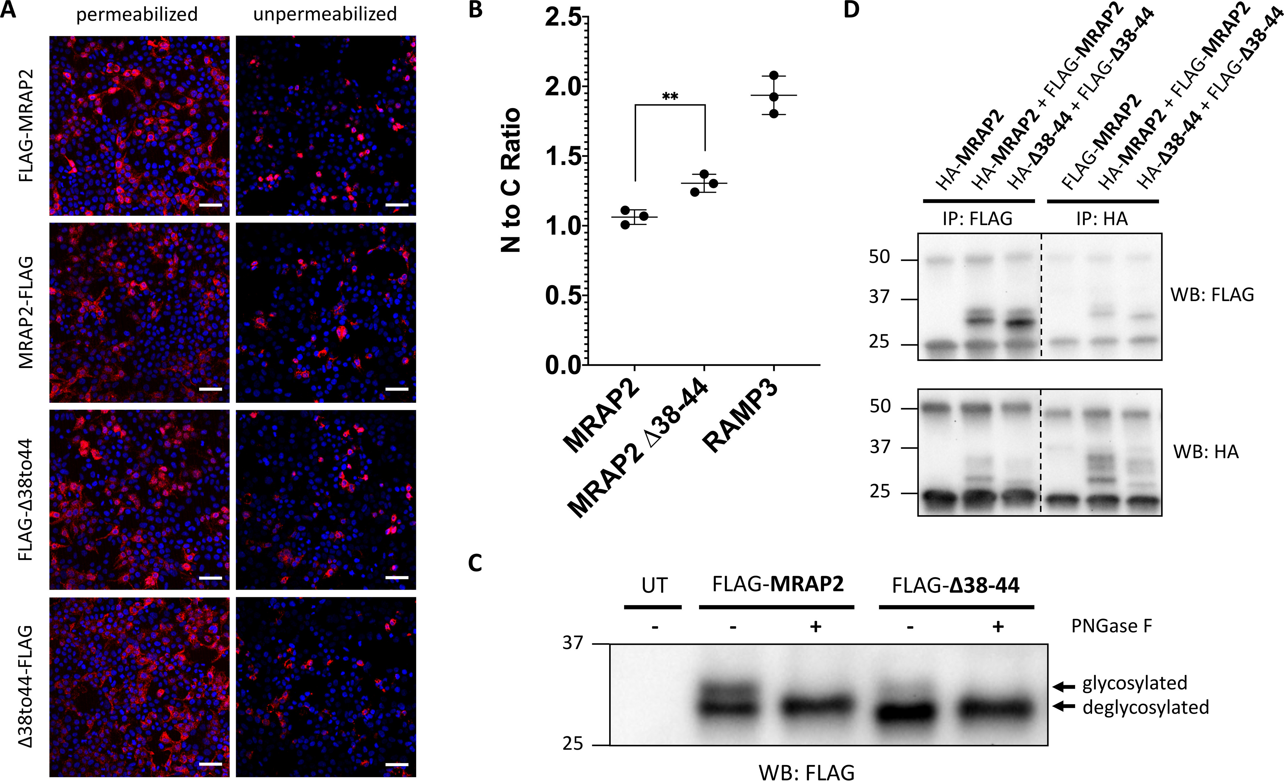
Figure 2.

The conserved motif required for dual topology and dimerization of MRAP1 is not required for dual topology and dimerization of MRAP2. A, both the N terminus and C terminus of MRAP2 WT and Δ38–44 are detected from intact, unpermeabilized HEK293T cells, as seen by immunofluorescence. FLAG-tagged MRAP2 is shown in pink, and the nucleus is shown in blue. Scale bars, 100 μm. B, flow cytometry was used to determine the N-terminal to C-terminal fluorescence ratio for MRAP2 WT, Δ38–44, and RAMP3 from intact cells expressing N-terminally tagged constructs and C-terminally tagged constructs. Expression levels for each construct were normalized using parallel experiments with permeabilized cells. The data represent the means from three independent experiments. Error bars show S.D. Statistical significance of differences was analyzed by t test. **, p < 0.01 versus MRAP2. C, immunoblot showing two protein species for FLAG-MRAP2 and FLAG-Δ38–44. Treatment with peptide:N-glycosidase F (PNGase F) results in a single, deglycosylated protein species. D, immunoblot showing co-immunoprecipitation of HA- and FLAG-tagged MRAP2 and HA- and FLAG-tagged Δ38–44. IP, immunoprecipitation; WB, Western blotting. UT, Untransfected.
