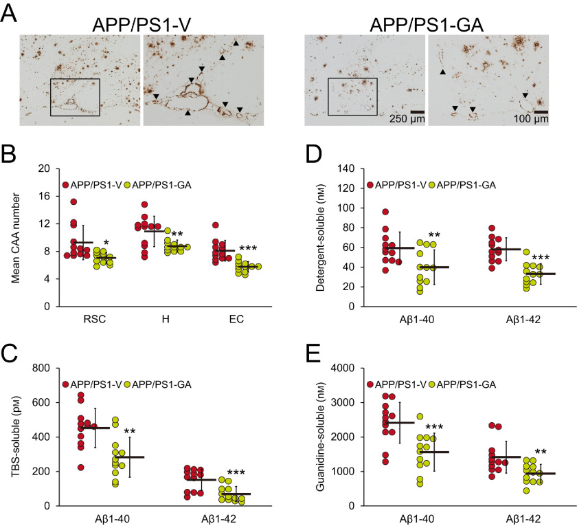
Figure 3.
GA therapy decreases cerebral vascular β-amyloid deposits and Aβ abundance. Representative images were obtained from vehicle- (APP/PS1-V) or GA- (APP/PS1-GA) treated APP/PS1 mice for 6 months starting at 12 months of age (mouse age at sacrifice = 18 months). A, immunohistochemistry using an Aβ17-24 mAb (4G8) highlights cerebral amyloid angiopathy as amber signals at the hippocampal fissure in the hippocampal area. Each image at right is a higher magnification image from insets. B, severity of cerebral amyloid angiopathy (mean numbers of CAA deposits per mouse) is shown on the y axis, and brain region is shown on the x axis (RSC, H, and EC). C–E, brain homogenates from TBS-soluble (C), detergent-soluble (D), or 5 m guanidine HCl-extractable (E) fractions were individually measured by sandwich ELISA for human Aβ1-40 and Aβ1-42. B–E, data were obtained from APP/PS1 mice treated with vehicle (APP/PS1-V, n = 12) or GA (APP/PS1-GA, n = 12) for 6 months beginning at 12 months of age (mouse age at sacrifice = 18 months). Data for B–E are presented as mean ± S.D. Statistical comparisons for B are between-groups within each brain region, and for C–E are between-groups for each Aβ species. *, p < 0.05; **, p < 0.01; ***, p ≤ 0.001 or p < 0.001 for APP/PS1-GA versus APP/PS1-V mice (B–E).

