Figure 2.

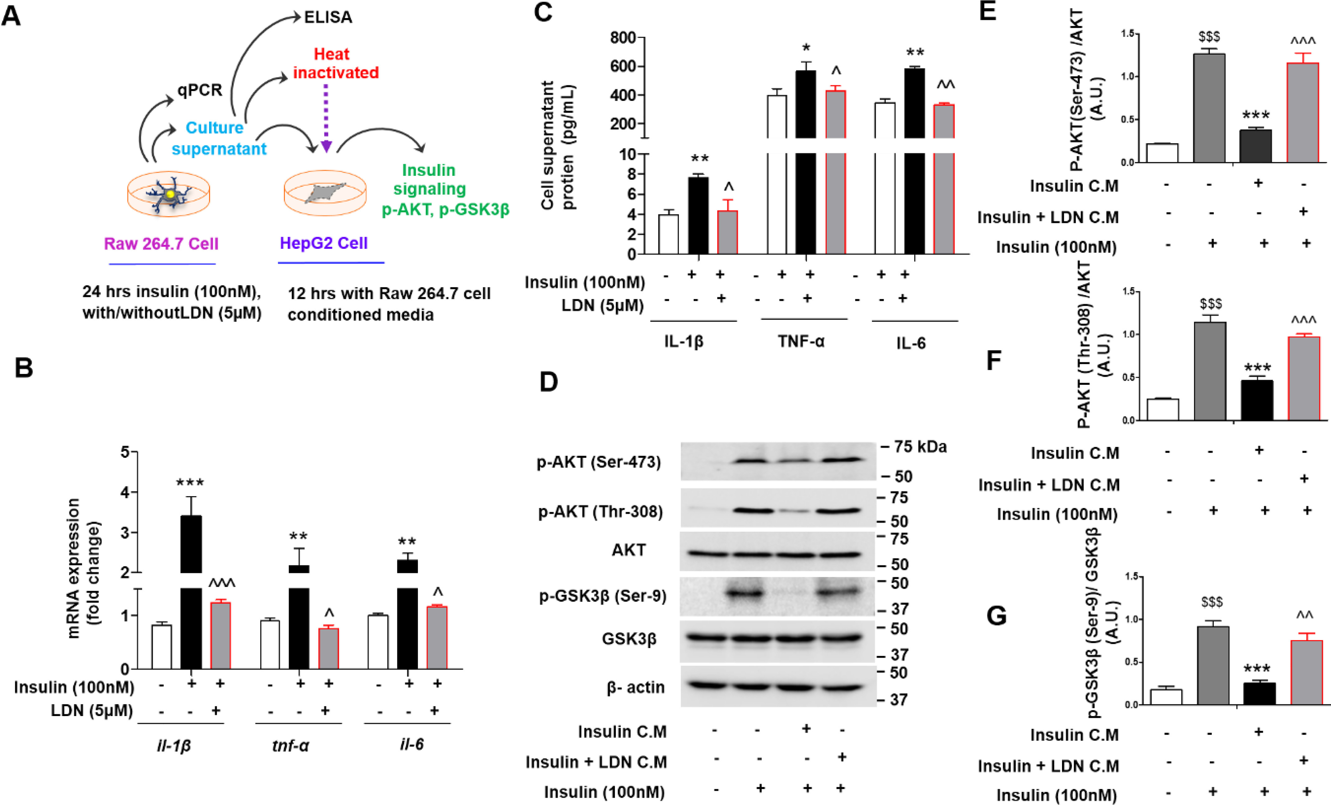
LDN inhibits inflammation and restored insulin sensitivity (surrogate markers AKT and GSK3β phosphorylation) in presence of hyperinsulinemia exposed conditioned media in vitro. A, schematic representing overall experimental design, and collection of conditioned media from murine macrophage (Raw 264.7) cells and treatment to hepatic (HepG2) cells. B and C, quantitative mRNA expression of indicated genes (il-1β, tnf-α, and il-6) (B) in murine macrophage cells, (C) levels of pro-inflammatory (IL-1β, TNFα, and IL-6) release in media from insulin challenged murine macrophage cells were measures using Bio-Plex ProTM mouse cytokine Standard 23 Plex Group 1 kit (Bio-Rad, 171304070M) on a Bio-Plex-200 (Bio-Rad). Values are expressed as mean ± S.D. ***, P < 0.001; **, P < 0.01; *, P < 0.05 versus control, and ^^^, P < 0.001; ^^, P < 0.01; ^, P < 0.05 versus insulin. (ANOVA followed by Bonferroni's Multiple Comparison). D–G, immunoblot (D) and (E–G) quantification for p-AKT (Ser-473), p-AKT(Thr-308), and p-GSK3β (Ser-9) phosphorylation status in HepG2 cell lysate treated with indicated conditioned media (C.M.) for 12 h followed by stimulated with or without 100 nm of insulin (last 15 min). Values are expressed as mean ± S.D. $$$P < 0.001 versus without insulin activation, ***, P < 0.001; **, P < 0.01; *, P < 0.05 versus insulin C.M. and ^^^, P < 0.001; ^^, P < 0.01; ^, P < 0.05 versus insulin + LDN C.M. $$$, P < 0.001; $$, P < 0.01; $, P < 0.05. (ANOVA followed by Bonferroni's Multiple Comparison).
