Figure 4.

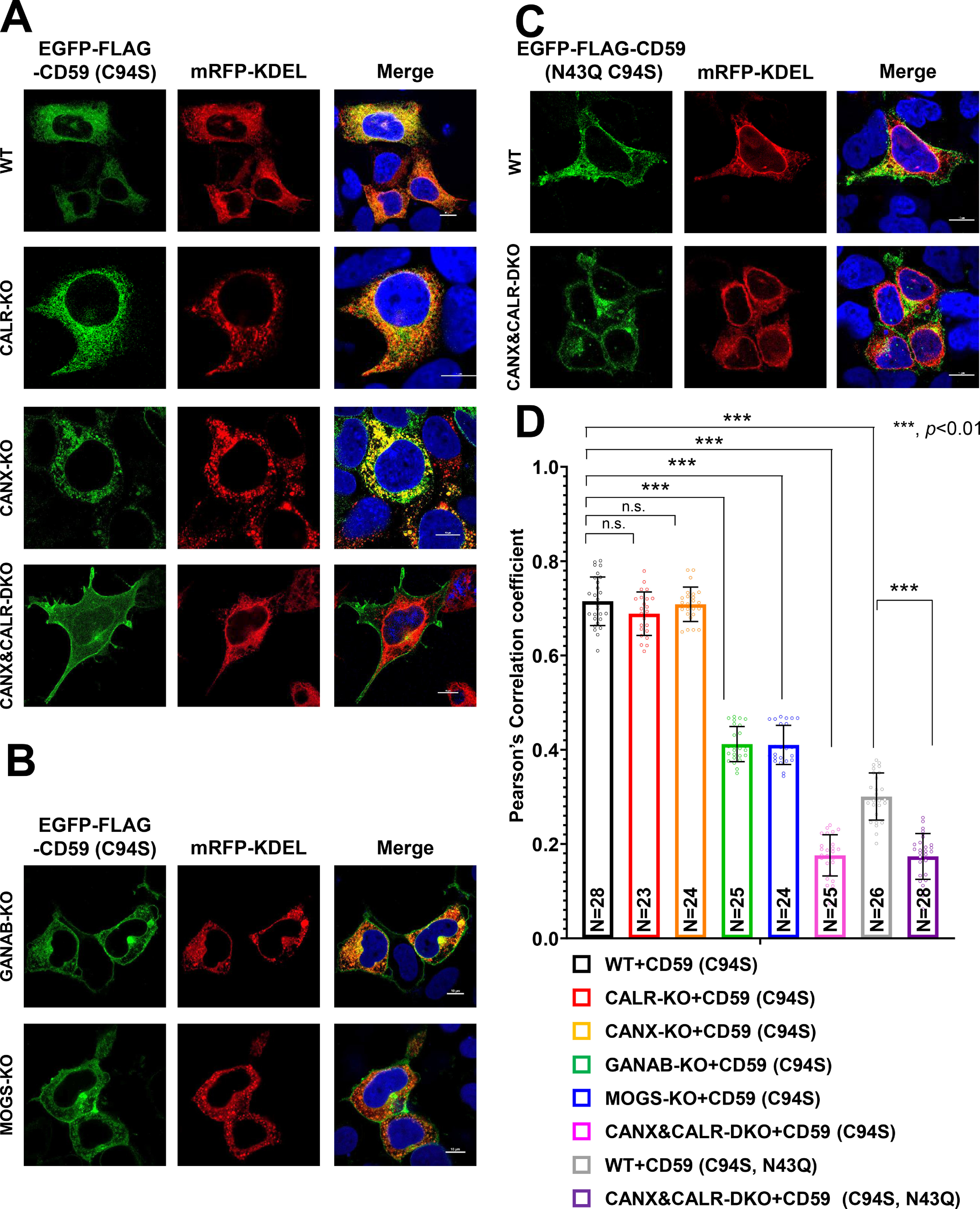
N-Glycan–dependent calnexin/calreticulin cycle mediates the efficient ER retention of misfolded GPI-APs. A–C, localization of misfolded CD59 (EGFP-FLAG-CD59 (C94S)) was analyzed in WT, CANX-KO, CALR-KO, and CANX&CALR-DKO cells (A) and in MOGS-KO and GANAB-KO cells (B). Localization of misfolded and non-N-glycosylated CD59 (EGFP-FLAG-CD59 (C94S, N43Q)) was performed as described in C. mRFP-KDEL was used as an ER marker. 3 days after transfection, images were obtained using confocal microscopy. DAPI staining is shown as blue in merged images. Scale bar, 10 μm. D, Pearson's correlation coefficient values between EGFP-FLAG-CD59 (C94S) or (C94S, N43Q) and mRFP-KDEL were calculated using the ImageJ plugin JACoP. The data are presented as the means ± S.D. (error bars) of the measurements. N, cell number used for the calculation. p values (two-tailed, Student's t test) are shown. n.s., not significant.
