Figure 8.

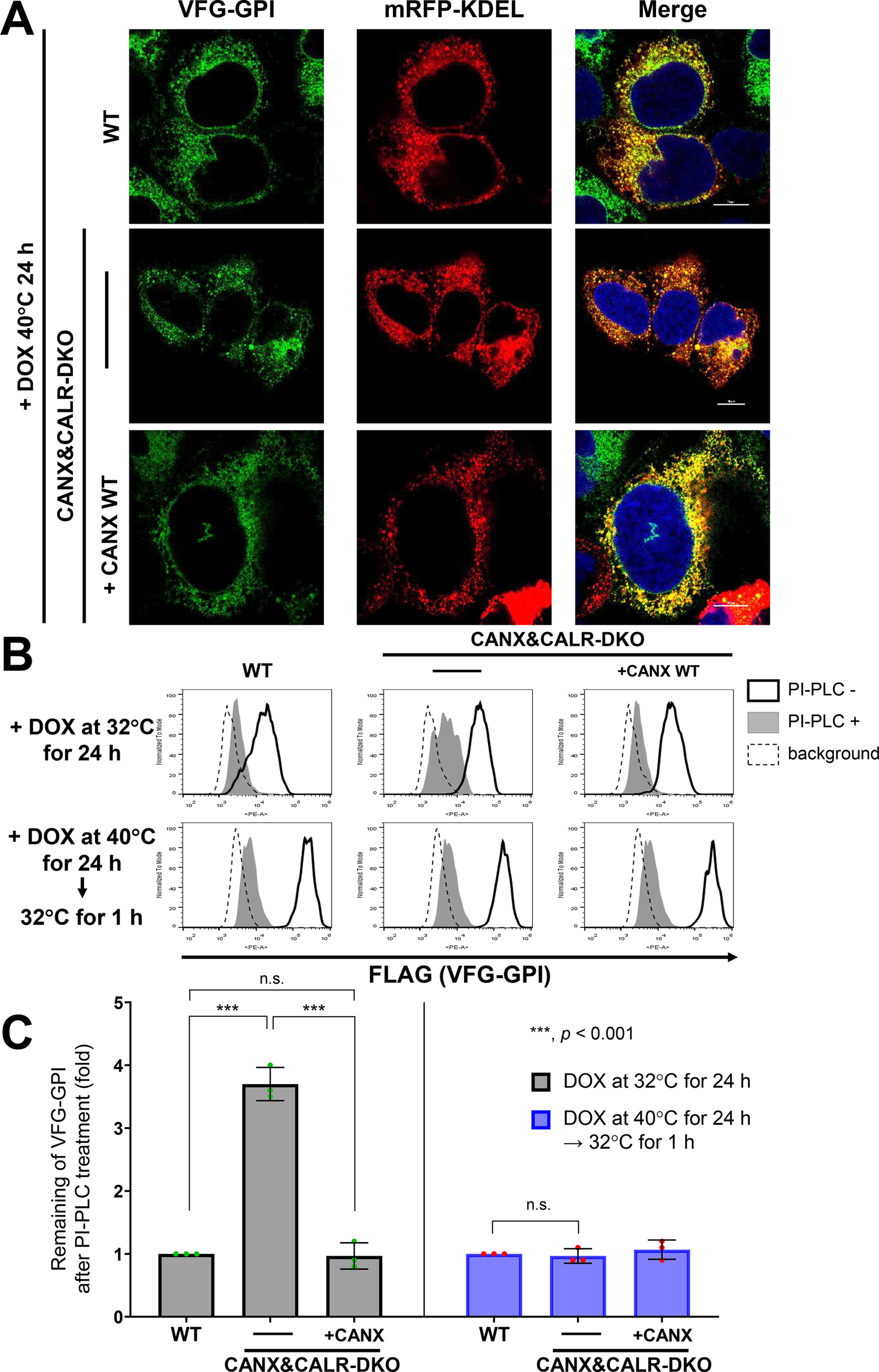
ER retention time regulates GPI-inositol deacylation. A, localization of VSVGts-FLAG-GFP-GPI (VFG-GPI) at 40 °C in WT, CANX&CALR-DKO, and CANX&CALR-DKO+CANX WT cells. The construct expressing mRFP-KDEL, an ER marker, was transfected into WT, CANX&CALR-DKO and CANX&CALR-DKO+CANX WT cells. At 36 h after transfection, cells were incubated with 1 µg/ml doxycycline at 40 °C for 24 h to induce VFG-GPI expression. Subsequently, the cells were quickly fixed with 4% paraformaldehyde and then imaged by confocal microscopy. Scale bar, 10 μm. B, HEK293FF6WT, CANX&CALR-DKO, and CANX&CALR-DKO+CANX WT cells were incubated with 1 µg/ml doxycycline at 32 °C for 24 h (top panels). Alternatively, cells were incubated with 1 µg/ml doxycycline at 40 °C for 24 h followed by a 32 °C incubation for 1 h (bottom panels). After induction, the cells were treated with or without PI-PLC, and surface VFG-GPI was stained with an anti-FLAG antibody and analyzed by flow cytometry. The region of same GFP intensity was gated to normalize VFG-GPI expression. C, remaining VFG-GPI after PI-PLC treatment in B was plotted. The values in WT cells were set as 1, and relative values in CANX&CALR-DKO were plotted. The data are presented as the means ± S.D. (error bars) of three independent measurements. p values (one-tailed, Student's t test) are shown. n.s., not significant.
