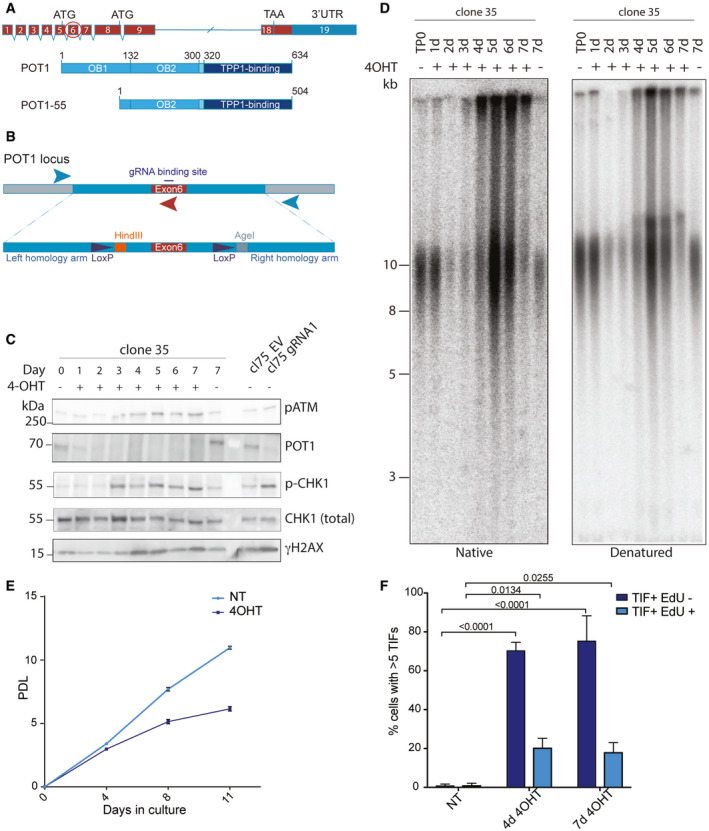
Figure 1. POT1‐loss triggers rapid telomere elongation and a DNA damage response throughout the cell cycle.

- Schematic drawing of the POT1 gene structure and two derived polypeptides. Exon 6 that has been chosen for gene editing is encircled.
- Schematic drawing of the repair template used for CRISPR/Cas9-mediated gene editing in HEK293E cells. Positions of primers for genotyping PCR are marked with arrows.
- Western blot for POT1 and DDR markers upon POT1 deletion induced with 0.5 μM 4‐OHT. Cl75—parental cell line. gRNA1—POT1 knockout in the population by transient transfection with pSpCas9(BB)‐2A-puro containing gRNA1 sequence. EV—corresponding empty vector.
- Time course of telomere length changes upon POT1 removal in clone 35. d (days) TP0—time point 0, no 4OHT.
- Growth curve for POT1 WT and POT1 knockout cells. Population doublings (PDL) are represented as mean ± SD (three biological replicates).
- Quantification for TIFs in EdU‐positive and EdU‐negative cells upon POT1 removal in clone 35. The bars show percentage of cells containing more than 5 TIFs ± SD. Experiments were performed in triplicate. At least 120 cells were analyzed per condition per replicate. Significance was determined using two‐way ANOVA. P‐values are indicated on the graph.
