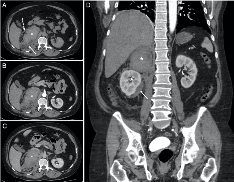
Figure 3.
Selected soft-tissue window (W:450, L:50) axial slices from the (A) non-contrast, (B) arterial and (C) venous phases of the abdomen and pelvis CT examination through the level of the coeliac trunk, with (D) venous phase coronal reconstruction, obtained the same day as the CTPA in figure 2. Images (A–C) confirm the recent right adrenal haemorrhage (*). The average density (HU, 55) is stable across all phases with no active extravasation of contrast. The haemorrhage extends to the right perirenal space (solid arrows in A and D) and tracks down into the right paracolic gutter, with thickening of the right anterior perirenal (Gerota) fascia (dashed arrow in A).

