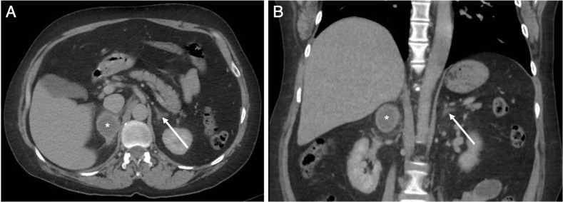
Figure 5.
Selected (A) axial and (B) coronal slices from a portal venous phase CT of the adrenal glands on soft-tissue window (W:450, L:50) obtained approximately 5 months after the CTPA (figure 2) and CT abdomen and pelvis (figure 3). The images demonstrate marked improvement in the appearances of the right adrenal gland (*) with subtotal resorption of the previous identified large adrenal haemorrhage (figures 2 and 3). There is no CT evidence of an underlying adrenal lesion and the left adrenal gland (arrow) is normal.

