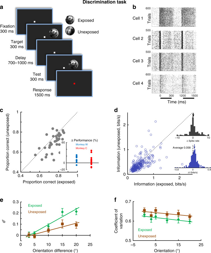
Fig. 3. Exposure improves neural coding in V1.
a Cartoon describing the discrimination task—monkeys are required to report whether two successively flashed images (target and test) differed or not in orientation (images were rotated by 0, 3, 5, 10, or 20°). Exposed and unexposed image trials were randomly interleaved (in 50% of trials the two images were not rotated with respect to each other). b Raster plots for example neurons recorded during image orientation discrimination task. The dark bars mark the stimulus presentation. c Proportion of total correct responses in the image discrimination task for the exposed vs. the unexposed image conditions. Each point is associated with a pair of images in one session (P = 0.00012, n = 34, Wilcoxon sign-rank test). Inset represents the change in perceptual performance in the exposed vs. unexposed conditions for each monkey. d Neurons transmit more information about the exposed images relative to unexposed images (P = 2.61 × 10⁻⁸, Wilcoxon sign-rank test, 300 ms, n = 263). Each point represents mutual information for a single neuron associated with the exposed (x axis) and unexposed (y axis) stimuli. (insets) (top) Distribution of firing rate elicited by the exposed and unexposed images. There are no significant changes in mean responses between the exposed and unexposed conditions. (bottom) Distribution of information differences, ΔI (Iexposed−Iunexposed) for the population of cells. e Mean neuronal sensitivity (d’) measured from the responses to the test stimuli (n = 263 neurons) is significantly greater for the exposed images (P = 0.0006, N-way ANOVA, df = 1, F = 11.94). f Mean coefficient of variation (CV) is lower for the exposed images compared with unexposed (P = 0.002, N-way ANOVA, df = 1, F = 9.53, n = 263 neurons). Error bars in all panels represent sem.

