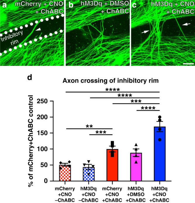
Fig. 1. Chemogenetic DRG neuron activation promotes neurite crossing of a ChABC-treated inhibitory proteoglycan barrier.

Representative images of DRG cultures growing on ChABC-treated aggrecan spots are shown in a–c. The inhibitory rim is demarcated in a. Quantification of the numbers of axons crossing the inhibitory rim is shown in d. Neurons and axons were visualized by βIII-tubulin staining (green). Without ChABC, neurons transduced with either AAV-mCherry or -hM3Dq failed to traverse the inhibitory rim. ChABC alone significantly enhanced axonal growth from both mCherry+ DRGs in the presence of CNO or hM3Dq+ DRGs in the absence of CNO. After ChABC digestion of CSPG, some axons from control, mCherry+ DRG neurons were able to grow across the inhibitory rim (a, arrowhead), similar to what we observed when hM3Dq+ DRG neurons are cultured in the absence of CNO (b, d). CNO-mediated activation of hM3Dq+ DRG neurons enabled more neurites to cross the inhibitory rim (arrows in c, d). More axons crossed the inhibitory rim when CNO-mediated chemogenetic activation of hM3Dq+ DRGs was combined with ChABC digestion of the inhibitory substrate. N = 16 spots/group. Mean ± SEM. One-way ANOVA and post-hoc multiple comparisons testing using the two-stage step-up method of Benjamini, Krieger, and Yekutieli, **p = 0.0054, ***p < 0.001 (hM3Dq+CNO-ChABC vs. mCherry+CNO + ChABC p = 0.0020; mCherry+CNO + ChABC vs. hM3Dq+CNO + ChABC p = 0.0003), ****p < 0.0001. Scale bar: 50 µm. Source data are provided as a Source Data file.
