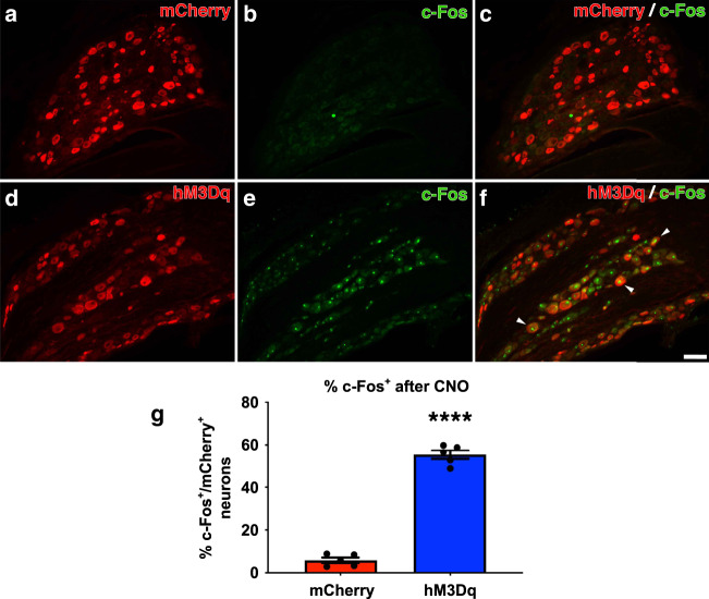
Fig. 3. CNO administration activates adult, hM3Dq+ DRG neurons in vivo.
AAV5-hSyn-hM3Dq-mCherry or AAV5-hSyn-mCherry was injected into the right C5-C8 DRGs. Four weeks later, CNO was subcutaneously injected into all animals. Two hours later, animals were perfused and DRG sections were stained for the mCherry reporter and c-Fos, an established marker of neuronal activation (a–f). Very few neurons transduced with AAV-mCherry expressed c-Fos (a–c) while many hM3Dq+ neurons expressed c-Fos (d–f, arrowheads). g Quantification of the percent of mCherry+ neurons that are also c-Fos+ after CNO administration. These data confirm that subcutaneous CNO injection chemogenetically activated hM3Dq+ neurons in vivo. N = 5 animals per group. Mean ± SEM. Two-tailed unpaired t-test, ****p < 0.0001. Scale bar: 100 µm. Source data are provided as a Source Data file.

