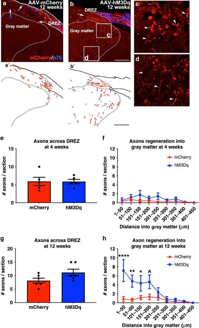
Fig. 6. Recurrent, chemogenetic activation of hM3Dq+ DRGs after a dorsal root crush promotes axon regrowth into the gray matter.
Transverse spinal cord tissue sections containing the dorsal root from animals 4 weeks or 12 weeks after dorsal root crush were immunostained for mCherry (red; to visualize axons from transduced DRG neurons) and p75 (blue; to visualize Schwann cells within the dorsal root to determine the boundary between the PNS and the CNS). Representative confocal images of the spinal cord sections with the dorsal root 12 weeks post-crush from mCherry+ animals (a) and hM3Dq+ animals (b–d) are shown. Traces of axon growth in the representative images of the mCherry+ and hM3Dq+ animals are shown in a’ and b’, respectively. Higher magnification images of regions in the section from the hM3Dq+ animal are shown in c and d. ChABC-digestion of CSPG at the DREZ enabled some axons to regenerate across the DREZ 4 weeks after dorsal root crush. There is no difference in the number of mCherry+ axons that regenerated across the DREZ (e) or into spinal gray ipsilateral to the crush (f) between mCherry+ or hM3Dq+ animals at this time point. As we saw 4 weeks post-crush, at 12 weeks post-crush, some axons extended across the ChABC-treated DREZ in both groups (a, b, g), including into white matter (arrows). Virtually no axons extended from control, mCherry+ DRGs into ipsilateral gray matter (a, a’, h). On the other hand, we saw more axons in ipsilateral dorsal horn from hM3Dq+ DRGs that received daily chemogenetic activation (arrowheads in c, d, h). N = 5 animals per group at 4 weeks. N = 6 animals per group at 12 weeks. Mean ± SEM. Two-way ANOVA and post-hoc multiple comparisons testing using the two-stage step-up method of Benjamini, Krieger, and Yekutieli, ^p = 0.0144, *p = 0.0199, **p = 0.0024, ****p = 0.0001. Scale bar: 75 µm. Source data are provided as a Source Data file.

