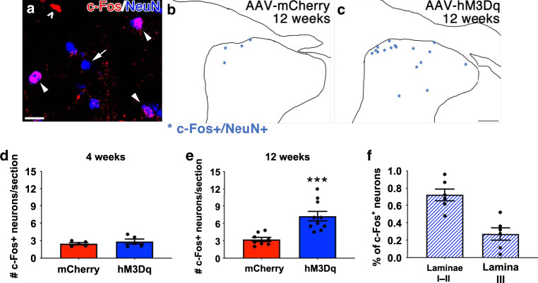
Fig. 7. Axons that regenerate into spinal gray matter upon chronic, CNO-induced chemogenetic neuron form functional synapses.
Four weeks and 12 weeks after complete C4-T1 dorsal root crush injuries, ipsilateral median and ulnar nerves were isolated and electrically stimulated for 30 min. Animals were sacrificed 1 hr later. Spinal cord sections between C5 and C8 were processed for immunohistochemistry to visualize c-Fos (red) and NeuN (blue), shown in a. Not all NeuN+ nuclei were also c-Fos+ (a, arrow) and not all c-Fos+ nuclei were in NeuN+ neurons (a, open arrowhead). Only c-Fos+/NeuN+ nuclei (a, closed arrowheads) were noted and counted. C-Fos+/NeuN+ nuclei in tracings of representative sections from mCherry- or hM3Dq-injected animals 12 weeks after crush are shown as asterisks in panels b and c. Four weeks after injury, electrical stimulation of the median and ulnar nerves induced c-Fos expression in few neurons within ipsilateral gray matter (d). There is no statistical difference between groups. At 12 weeks post-injury, more c-Fos+ neurons were observed in gray matter in animals that received repeated, daily activation of hM3Dq+ DRGs (e). The majority of these c-Fos+ neurons were located in more superficial laminae I and II (72.1 ± 0.7%). However, an appreciable percentage (27.3 ± 0.7%) was present in lamina III (f). N = 5 animals per group at 4 weeks; N = 8 mCherry+ animals and 10 hM3Dq+ animals at 12 weeks. Mean ± SEM. Two-tailed unpaired t-test, ***p = 0.0008. Scale bars: a 10 µm; c 100 µm. Source data are provided as a Source Data file.

